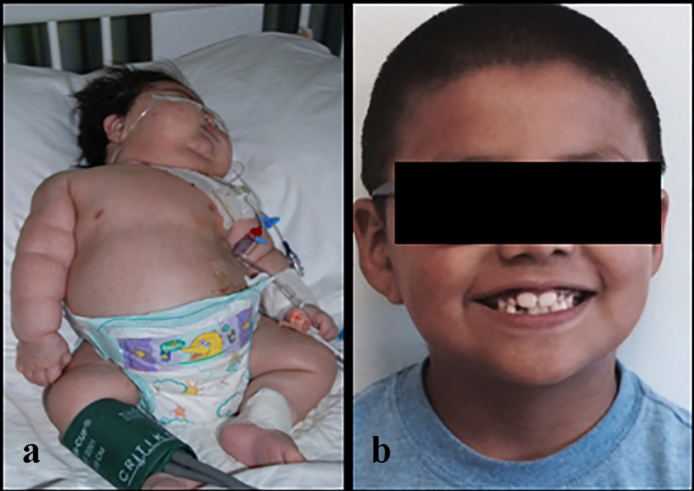
Figure 1. A 4-month-old male infant with an adrenocortical carcinoma at the time of presentation (a) and the same boy (b) at 7 years of age.
| World Journal of Oncology, ISSN 1920-4531 print, 1920-454X online, Open Access |
| Article copyright, the authors; Journal compilation copyright, World J Oncol and Elmer Press Inc |
| Journal website http://www.wjon.org |
Case Report
Volume 8, Number 3, June 2017, pages 81-85
Rare Presentation of Adrenocortical Carcinoma in a 4-Month-Old Boy
Figures
Table
| Author | Age at diagnosis | Sex | Presentation |
|---|---|---|---|
| Fudge et al [4] | 6 months | F | Isolated Cushing’s |
| Garge et al [5] | 3 months | M | Isolated Cushing’s |
| De Leon et al [6] | 2 months | M | Isolated Cushing’s |
| Romaguera et al [7] | 4 years | F | Cushingoid features, virilization |
| Kanmaz et al [8] | 4 years | F | Abdominal pain, non-functional tumor |
| Breidbart et al [9] | 3 years and 5 months | F | Virilization |
| Kim et al [10] | 8 years and 2 months | M | Virilization, peripheral precocious puberty |
| Arico et al [11] | 2 years | F | Virilization |
| Wong et al [12] | 12 years | F | Secondary amenorrhea, virilization, Cushing’s syndrome |
| Sorgo et al [13] | 12 years and 10 months | F | Virilization |
| Ghazizadeh et al [14] | 2 years | F | Virilization, heterosexual pseudoprecocious puberty |