Figures
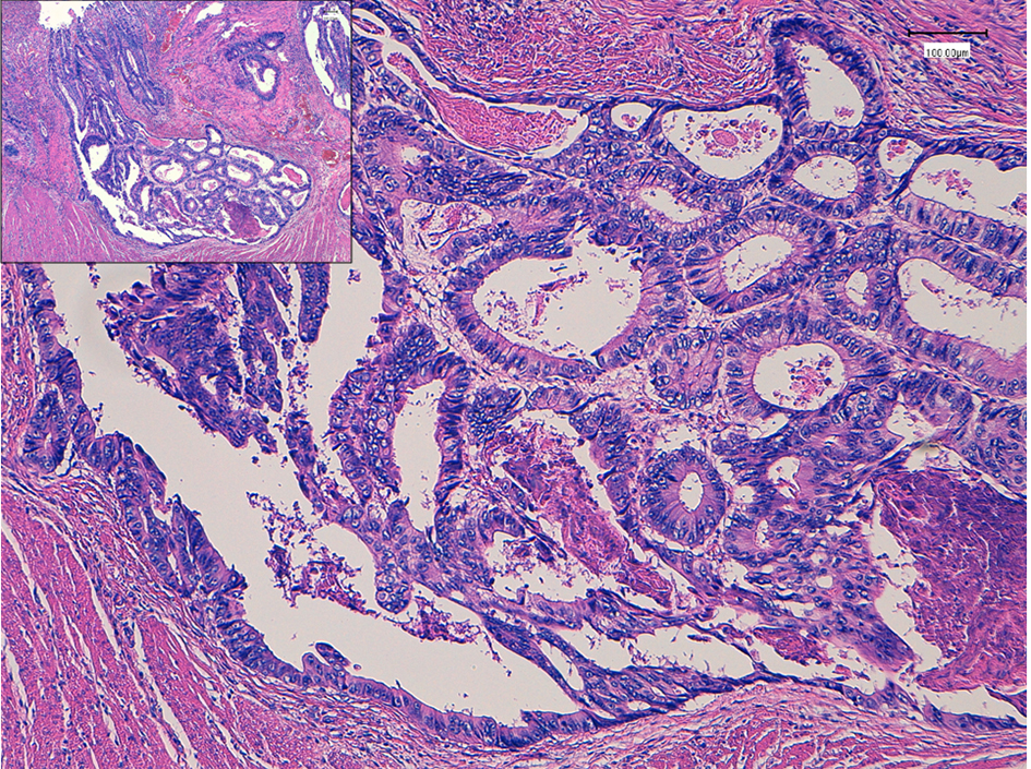
Figure 1. Histopathology of colon cancer, which was observed in the sigmoid colon 7 years ago. Moderately differentiated adenocarcinoma (tub2) invaded into the muscular layer. Insert is a low power of view. Bars, 100 μm. Hematoxylin and eosin stain.
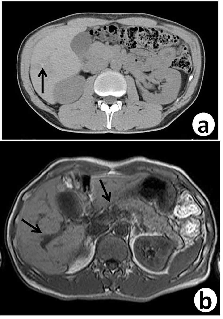
Figure 2. Follow-up abdominal contrast-enhanced CT scan (a) and MRI-T2 (b), 7 years after sigmoidectomy, reveal small dilated intrahepatic bile ducts (arrows) with thickening in the S5/6 area.
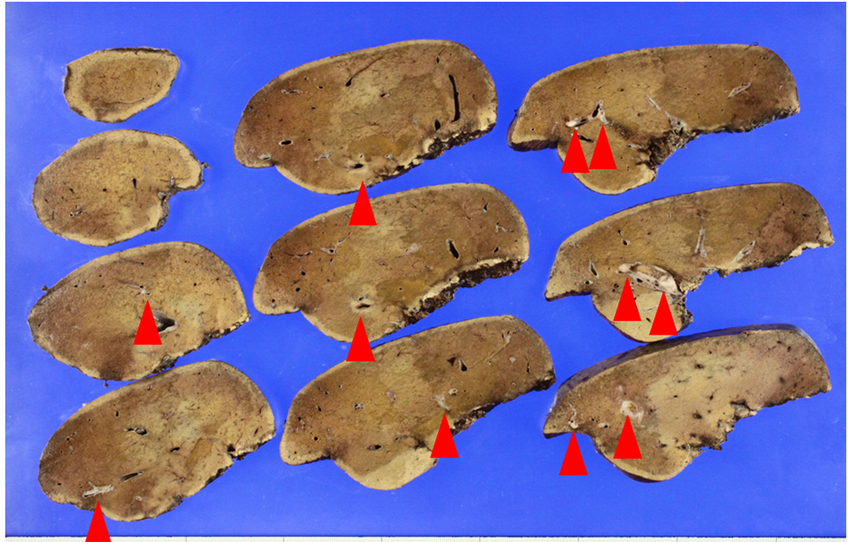
Figure 3. Macroscopic view of the lobectomized liver. Metastatic nodules (arrows) are found along with intrahepatic bile ducts.
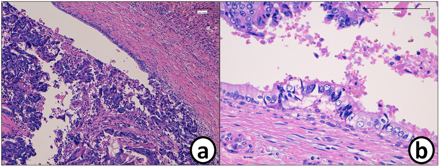
Figure 4. (a) Histopathology of the metastatic tumor in the liver (S5/6). (b) Mass of the cancer cells in the bile duct. Cancer cell mass is attached to the bile duct epithelium. A small metastatic focus is detected in the bile duct epithelium. Bars, 100 μm. Hematoxylin and eosin stain.

Figure 5. Metastatic cancer cells in the bile duct are positive for (a) CDX2 and (b) p53, while they are negative for (c) CK7 and (d) CK20. Bars, 100 μm. Immunohistochemistry for (a) CDX2, (b) p53, (c) CK7 and (d) CK20.
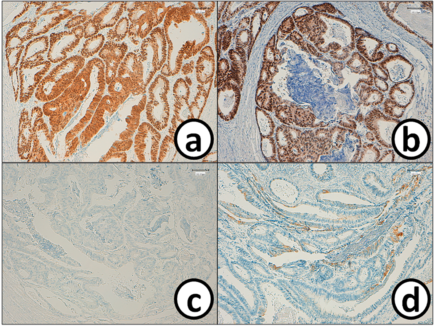
Figure 6. Cancer cells in the sigmoid colorectum are positive for (a) CDX2 and (b) p53, while they are negative for (c) CK7 and (d) CK20. Bars, 100 μm. Immunohistochemistry for (a) CDX2, (b) p53, (c) CK7 and (d) CK20. Bars, 100 μm.
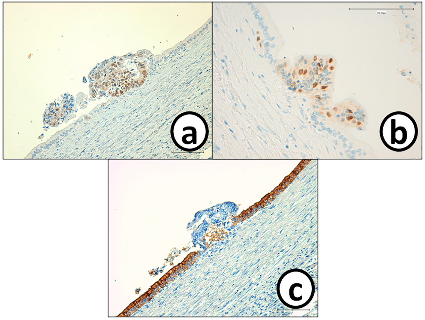
Figure 7. Cancer cells in the small metastatic focus in the bile duct epithelium are p53 and MIB-1, but negative for CA19-9. On the other hand, bile ducts epithelium is negative for (a) p53 and (b) MIB-1, but positive for (c) CA19-9. Immunohistochemistry of (a) p53, (b) MIB-1 and (c) CA-19-9. Bars, 100 μm.
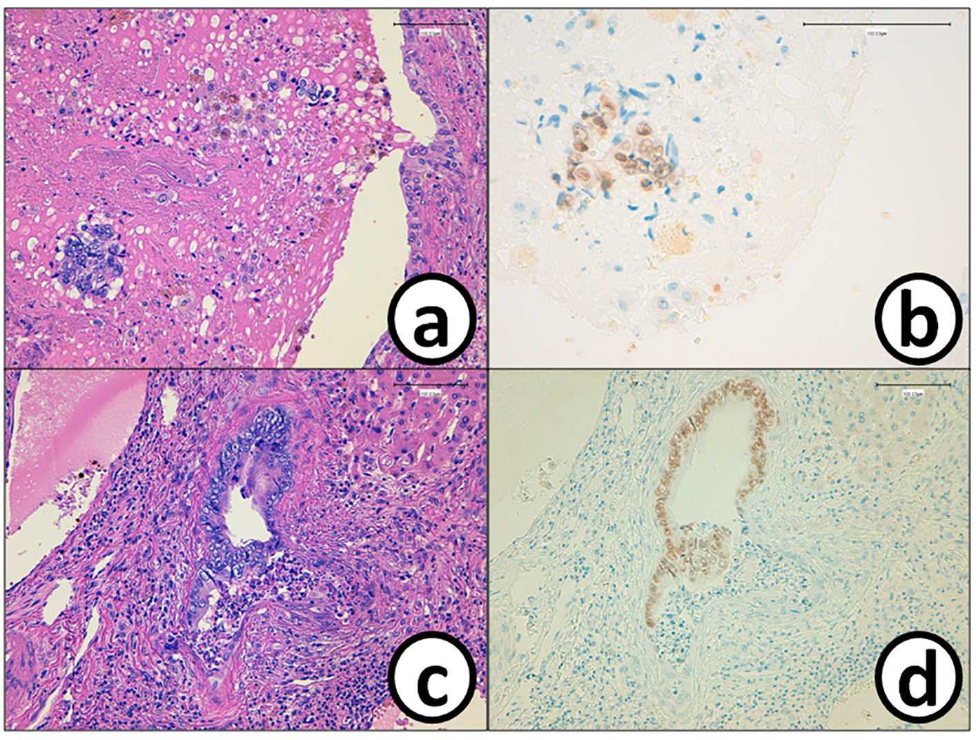
Figure 8. Cancer cells (a) in the tumor thrombus in the bile duct are positive for (b) CDX2. The epithelium of the bile duct is replaced by (c) metastatic colon cancer cells with (d) positive reaction of CDX2. Bars, 100 μm. (a, c) Hematoxylin and eosin stain. (b, d) Immunohistochemistry of CDX2.
Table
Table 1. Comparison of Immunohistochemical Findings
| Antibodies | Cancer cells in | Intrahepatic bile duct epithelium |
|---|
| Colon | Liver |
|---|
| CK7 | Negative (Fig. 6c) | Negative (Fig. 5c) | Positive (Fig. 5c) |
| CK17 | Positive | Positive (partial) | Positive (partial) |
| CK19 | Positive | Positive (partial) | Positive |
| CK20 | positive (partial) (Fig. 6d) | Negative (Fig. 5d) | Negative (Fig. 5d) |
| CDX2 | Positive (Fig. 6a) | Positive (Fig. 5a) | Negative (Fig. 5a) |
| MUC-5AC | Negative | Negative | Negative |
| MUC2 | Negative | Negative | Negative |
| P53 | Positive (Fig. 6b) | Positive (Fig. 5b) (Fig. 7a) | Negative |
| CA19-9 | Negative | Negative (Fig. 7c) | Positive (Fig. 7c) |
| MIB-1 positive rate | 50% | 30% (Fig. 7b) | 0% (Fig. 7b) |