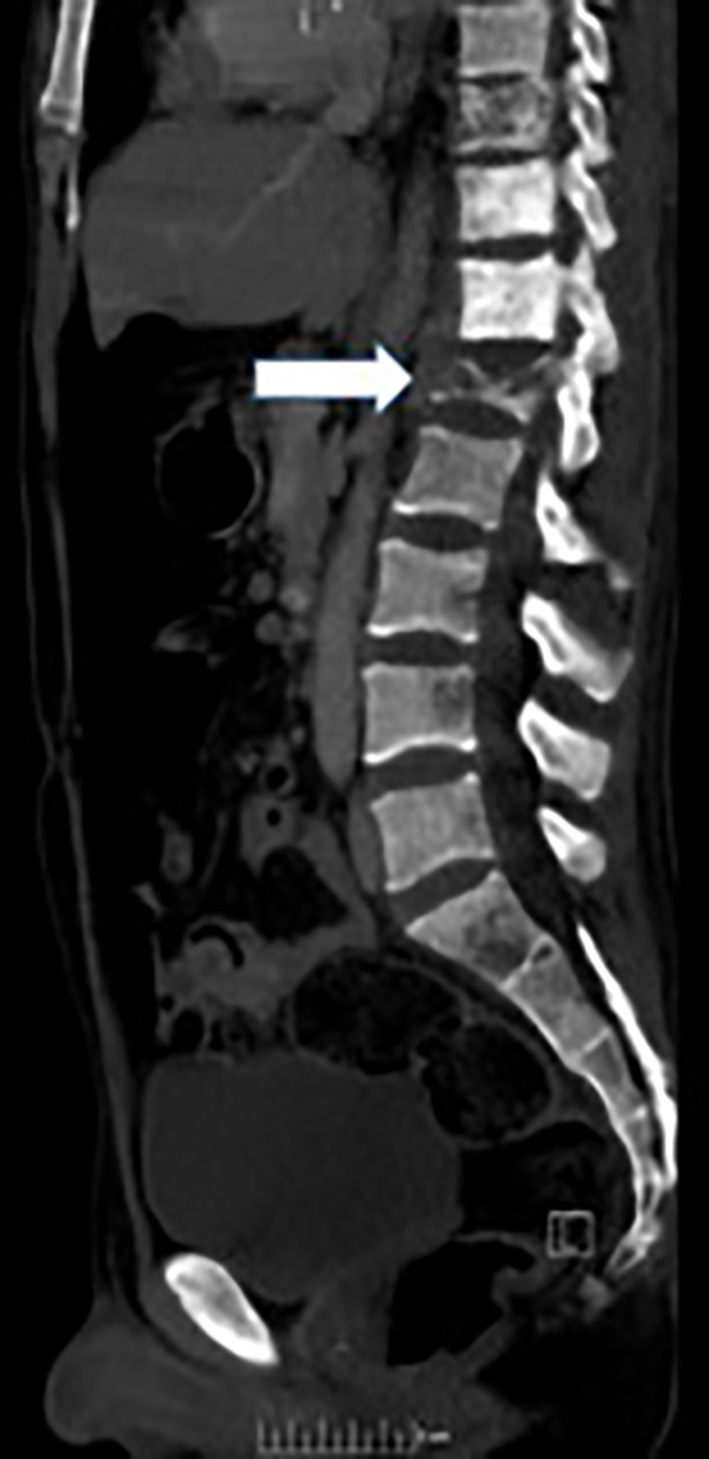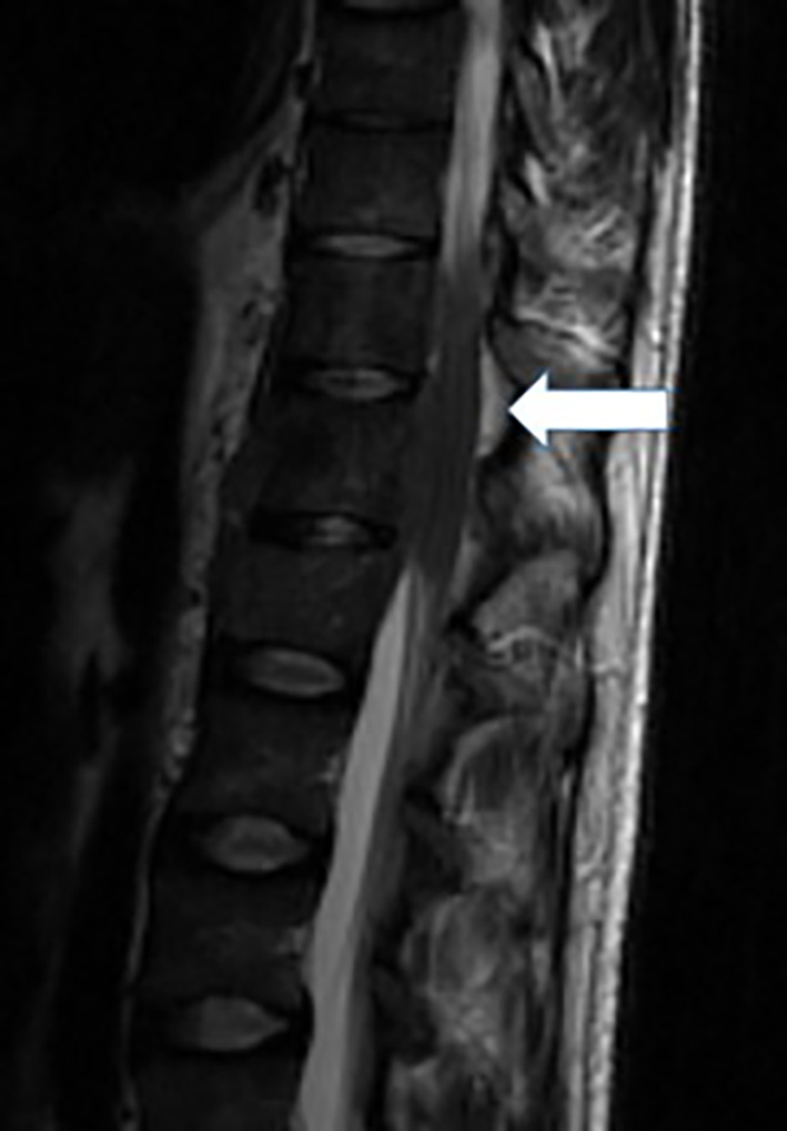
Figure 1. MRI spine depicting the lytic destruction of the spinal bodies and epidural mass at level T12-L1.
| World Journal of Oncology, ISSN 1920-4531 print, 1920-454X online, Open Access |
| Article copyright, the authors; Journal compilation copyright, World J Oncol and Elmer Press Inc |
| Journal website http://www.wjon.org |
Case Report
Volume 8, Number 4, August 2017, pages 122-125
Rare Case of Spinal Cord Compression as Initial Presentation of Thymic Carcinoma
Figures