Figures
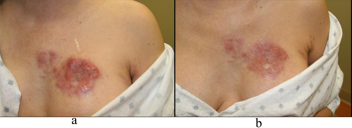
Figure 1. a: Left chest nodules; b: Left chest nodules.
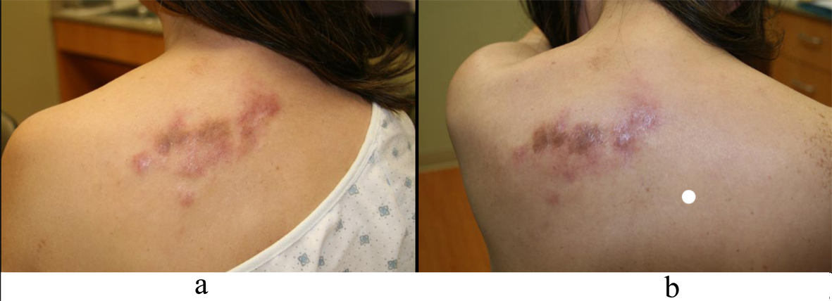
Figure 2. a: Left back nodules; b: Left back nodules.
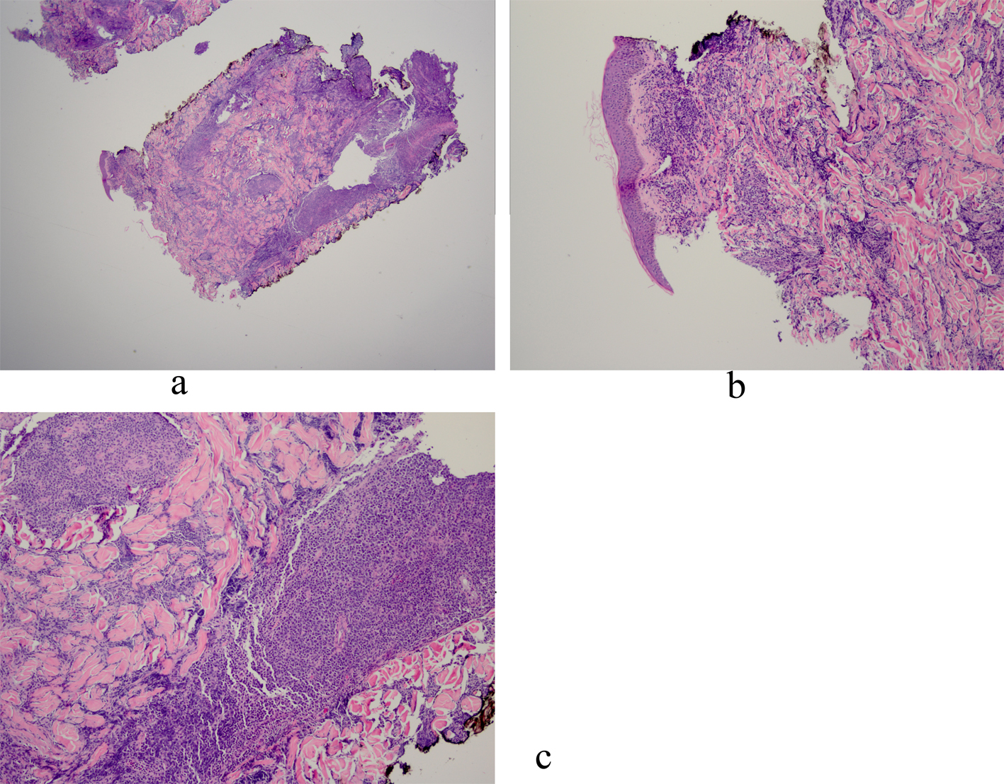
Figure 3. a: Biopsy of left chest nodule reveals atypical lymphoid infiltrate in the dermis with no infiltration into the epidermis; b: Biopsy of left chest nodule reveals atypical lymphoid infiltrate in the dermis with no infiltration into the epidermis; c: Biopsy of left chest nodule reveals atypical lymphoid infiltrate in the dermis with no infiltration into the epidermis.
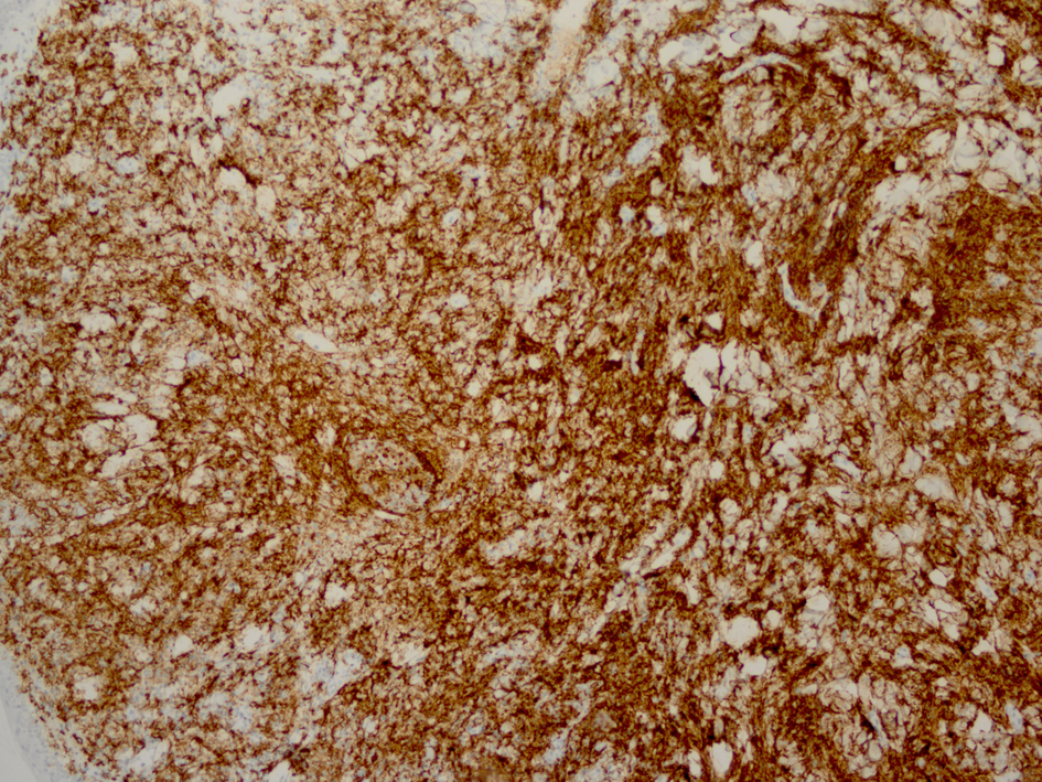
Figure 4. Immunostaining reveals monomorphic atypical lymphocytes diffusely arranged through the dermis as CD20 positive B-cells.
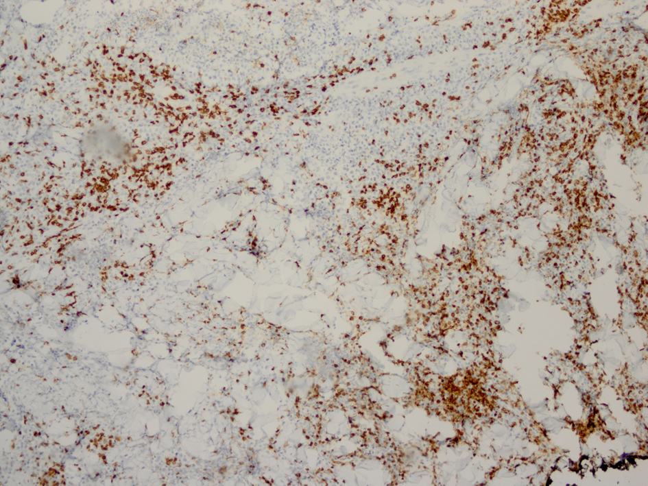
Figure 5. Immunostaining for CD3 reveals smaller non-atypical cells, consistent with reactive T-cells.
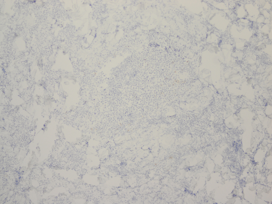
Figure 6. Immunostaining for CD30 reveals few scattered reactive cells.
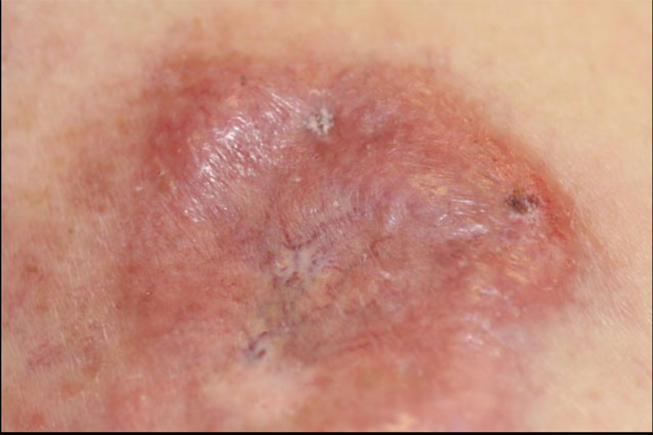
Figure 7. Left breast and inferior left chest wall with skin thickening and nodularity.