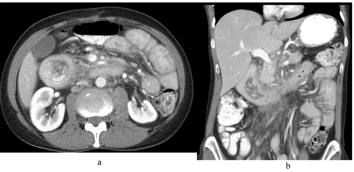
Figure 1. CT scan of abdomen and pelvis with IV contrast (A) axial view and (B) coronal view showing marked distention with irregular wall thickening of the duodenum just proximal to the genu causing a partial gastric outlet obstruction.
| World Journal of Oncology, ISSN 1920-4531 print, 1920-454X online, Open Access |
| Article copyright, the authors; Journal compilation copyright, World J Oncol and Elmer Press Inc |
| Journal website http://www.wjon.org |
Case Report
Volume 4, Number 2, April 2013, pages 102-106
Recurrent Adenocarcinoma of Colon Presenting as Duodenal Metastasis With Partial Gastric Outlet Obstruction: A Case Report With Review of Literature
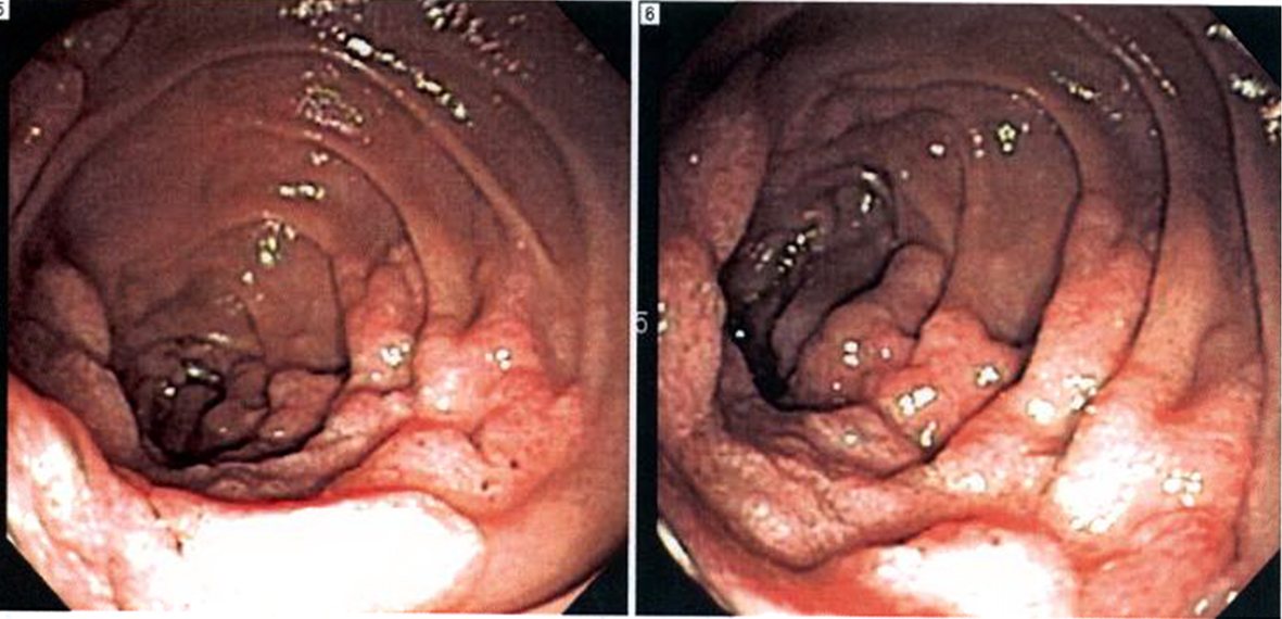
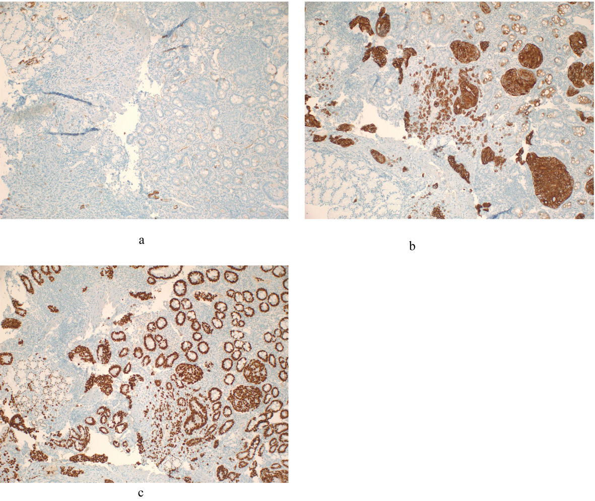
Figures