World Journal of Oncology, ISSN 1920-4531 print, 1920-454X online, Open Access
Article copyright, the authors; Journal compilation copyright, World J Oncol and Elmer Press Inc
Journal website http://www.wjon.org

Case Report

Volume 4, Number 3, June 2013, pages 161-164


Malignant Peripheral Nerve Sheath Tumors Masking as Ewing Sarcoma/Primitive Neuroectodermal Tumors

Figures

Figure 1.
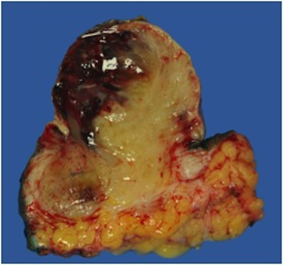
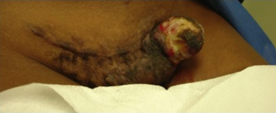
Figure 1. Gross unresected tumor on lateral side of C-section incision.
Figure 2.
Figure 2. Pelvic CT with abdominal wall soft tissue stranding after tumor resection.
Figure 3.
Figure 3. PET CT with hypermetabolic activity with abdominal wall soft tissue stranding and pelvic/inguinal hypermetabolic lymphadenopathy.
Figure 4.
Figure 4. Gross resected tumor with fatty and myxoidal features and focal hemorrhage.
Figure 5.
Figure 5. 20 × view with spindle cells and areas with loose and dense matrix of small round blue cells.
Figure 6.
Figure 6. 40 × view with small round blue cells as well as spindle cells arranged in a wavy fasicular pattern suggestive of neural origin.