World Journal of Oncology, ISSN 1920-4531 print, 1920-454X online, Open Access
Article copyright, the authors; Journal compilation copyright, World J Oncol and Elmer Press Inc
Journal website http://www.wjon.org

Case Report

Volume 4, Number 4-5, October 2013, pages 201-204


Management of Metastatic Solid Pseudopapillary Cancer of the Pancreas: A Case Report

Figures

Figure 1.
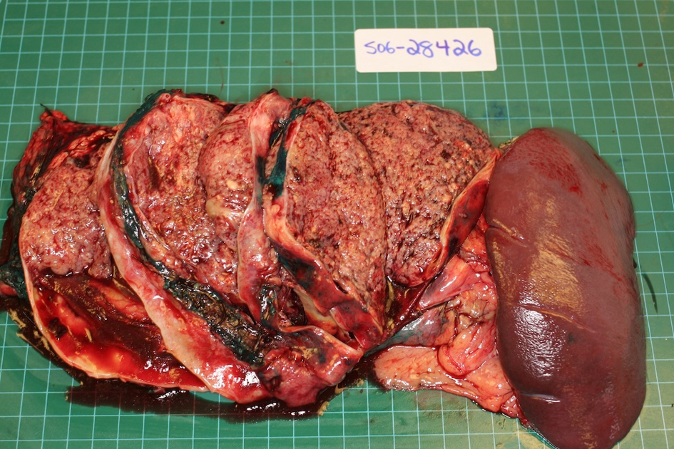
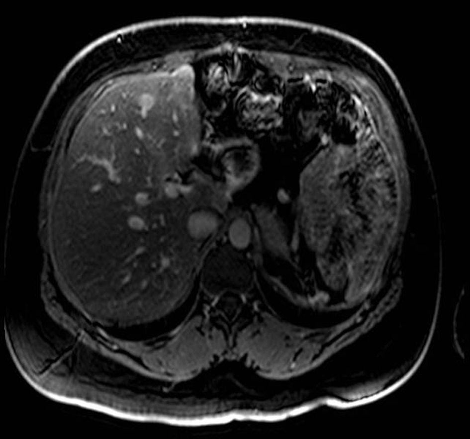
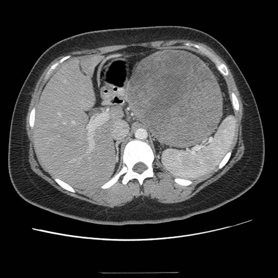
Figure 1. CT scan of the abdomen demonstrating a large mass arising from the tail of the pancreas.
Figure 2.
Figure 2. Surgical specimen demonstrating large tumor in the tail of the pancreas with prominent cystic degeneration.
Figure 3.
Figure 3. Low power micrograph showing normal pancreas (right) and tumor (left). Note cholesterol clefts in tumor.
Figure 4.
Figure 4. MRI with gadolinium contrast demonstrating a metastases in segment 4 of the liver.
Figure 5.
Figure 5. Micrograph of one of the liver metastases. The tumor demonstrates neoplastic cells supported by delicate vessels, pseudopapillary growth pattern, and cholesterol clefts.