World Journal of Oncology, ISSN 1920-4531 print, 1920-454X online, Open Access
Article copyright, the authors; Journal compilation copyright, World J Oncol and Elmer Press Inc
Journal website http://www.wjon.org

Case Report

Volume 5, Number 2, April 2014, pages 72-76


Neuroendocrine Differentiation of Prostate Cancer Metastases Evidenced “in Vivo” by 68Ga-DOTANOC PET/CT: Two Cases

Figures

Figure 1.
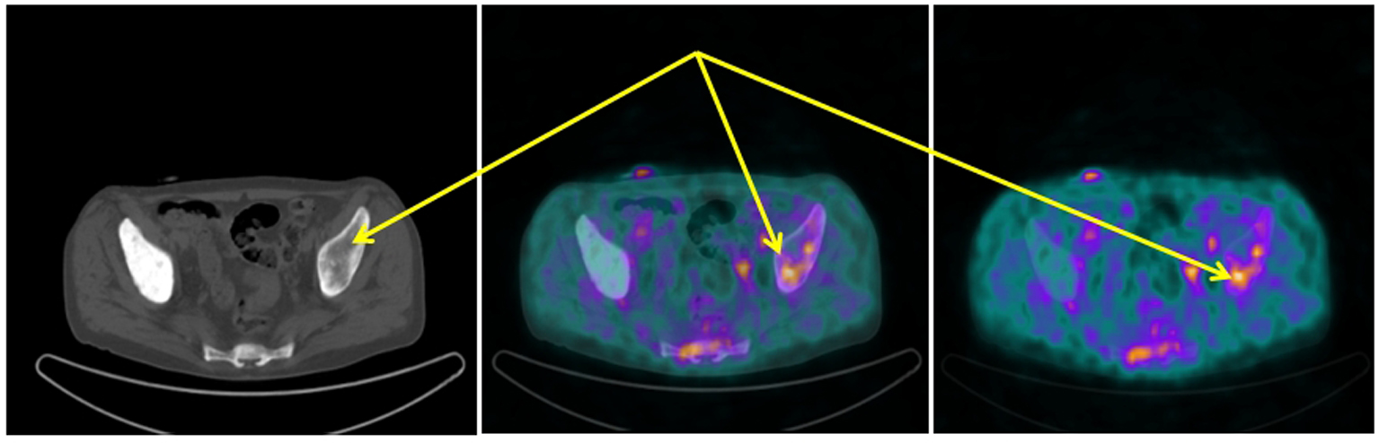
Figure 1. 68Ga-DOTANOC transaxial PET/CT (left CT attenuation correction, middle fused PET/CT, right PET only) showing increased uptake of the radiopharmaceutical in the left sovra-acetabular region corresponding to a mixed (predominantly lytic) skeletal metastasis (arrows).
Figure 2.
Figure 2. Same patient and acquisition parameters of Fig. 1. 68Ga-DOTANOC transaxial PET/CT (left CT attenuation correction, middle fused PET/CT, right PET only) showing increased uptake corresponding to lymphangitic carcinomatosis of the right lung (arrows).
Figure 3.
Figure 3. 68Ga-DOTANOC transaxial PET/CT at the level of left humeral head showing increased uptake of the radiopharmaceutical in a mixed metastasis (pointers).
Figure 4.
Figure 4. Same patient and acquisition parameters of Fig. 3. 68Ga-DOTANOC transaxial PET/CT at the level of fourth lumbar vertebra showing increased uptake of the radiopharmaceutical in a mixed metastasis.