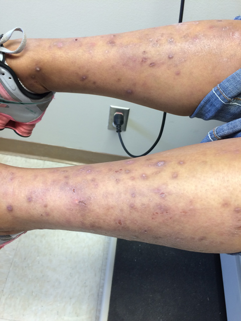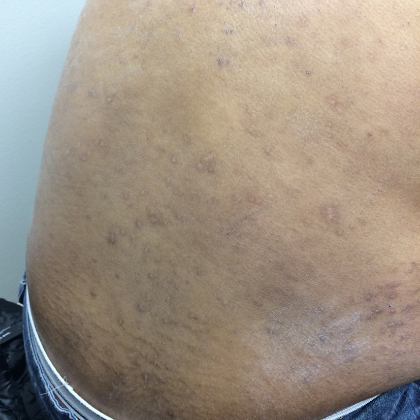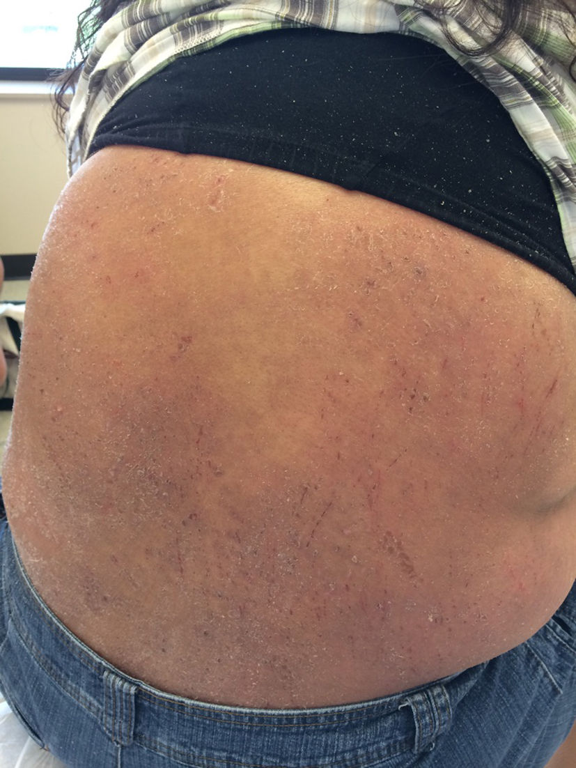
Figure 1. Excoriation and lesions on patient’s flank on presentation.
| World Journal of Oncology, ISSN 1920-4531 print, 1920-454X online, Open Access |
| Article copyright, the authors; Journal compilation copyright, World J Oncol and Elmer Press Inc |
| Journal website http://www.wjon.org |
Case Report
Volume 6, Number 2, April 2015, pages 335-337
Langerhan Cell Histiocytosis: A Rare Disorder With a Rare Presentation
Figures