World Journal of Oncology, ISSN 1920-4531 print, 1920-454X online, Open Access
Article copyright, the authors; Journal compilation copyright, World J Oncol and Elmer Press Inc
Journal website http://www.wjon.org

Case Report

Volume 6, Number 3, June 2015, pages 383-386


Gastric Perforation in a Patient Receiving Neoadjuvant Chemoradiotherapy

Figures

Figure 1.
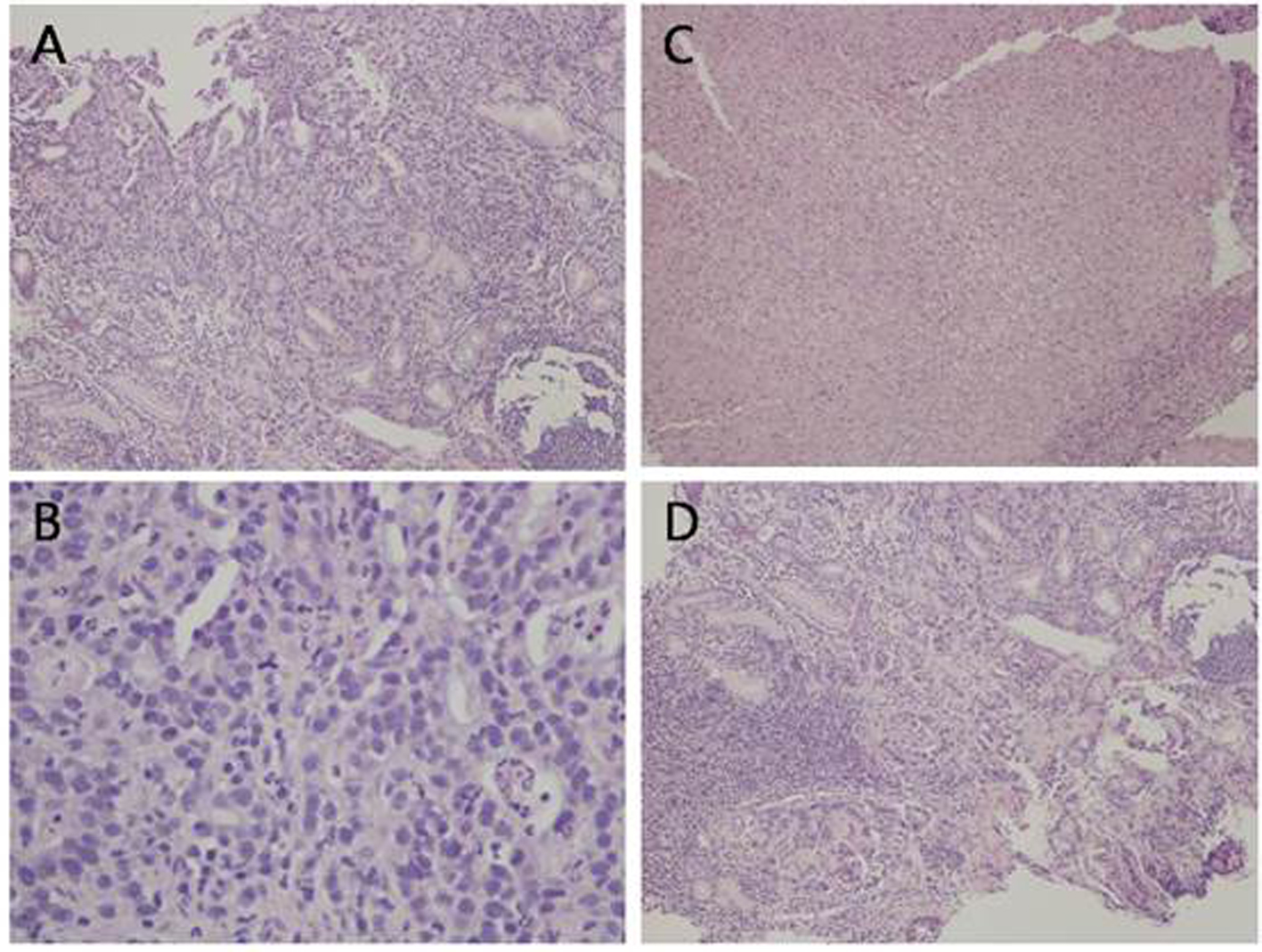
Figure 1. Histological analysis of the gastric ulcer biopsy showing malignant glandular proliferation (A, H&E, × 10), tumor cells exhibiting high nuclear to cytoplasmic ratio, prominent nucleoli, and frequent mitotic figures (B, H&E, × 40). There are areas of ulceration with tissue necrosis (C, H&E, × 10) and areas of lymphocytic infiltrate, morphologically consistent with reactive process (D, H&E, × 10).
Figure 2.
Figure 2. CT showing moderate pneumoperitoneum (arrow) and perihepatic fluid (arrow head).