World Journal of Oncology, ISSN 1920-4531 print, 1920-454X online, Open Access
Article copyright, the authors; Journal compilation copyright, World J Oncol and Elmer Press Inc
Journal website http://www.wjon.org

Case Report

Volume 7, Number 1, February 2016, pages 21-27


Desmoid Tumors: A Clear Perspective or a Persisting Enigma? A Case Report and Review of Literature

Figures

Figure 1.
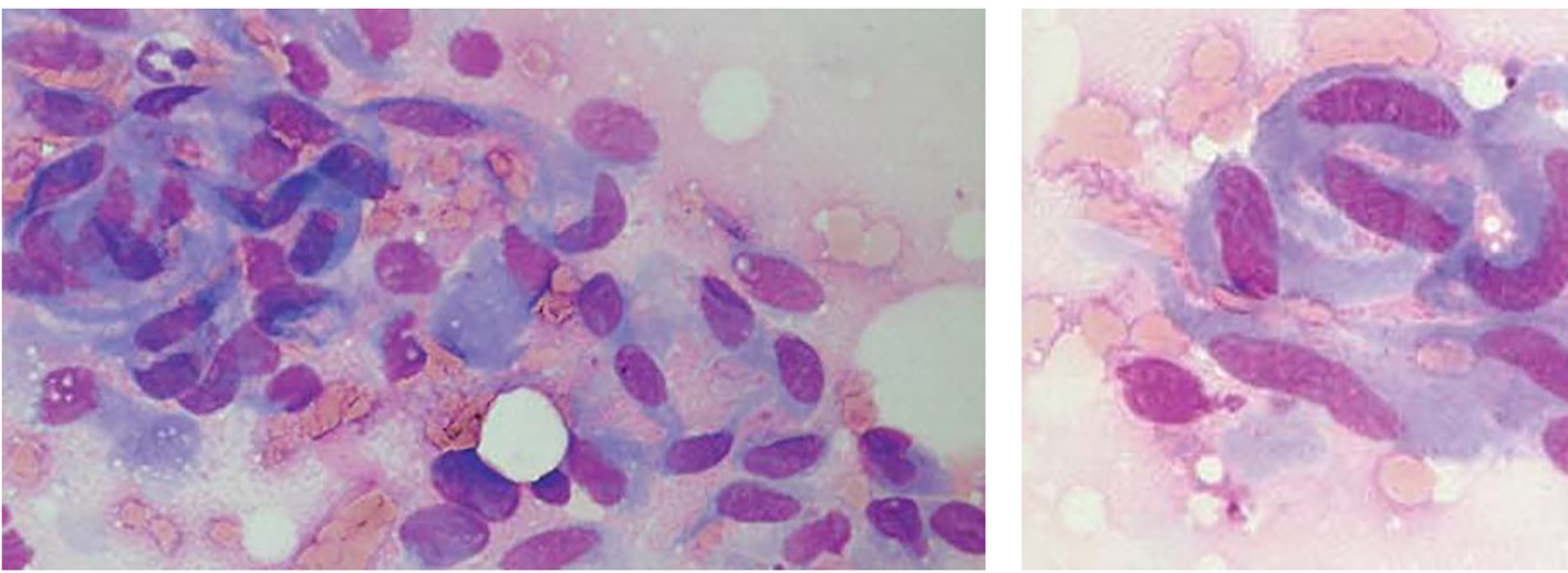
Figure 1. Fine-needle aspirates of desmoid tumors showing coherent clusters of uniform spindle cells with abundant cytoplasm and oval to elongated nuclei with evenly distributed chromatin. Large, basophilic multinucleate cells representing atrophic muscle fibers are also seen.
Figure 2.
Figure 2. Core needle biopsies of desmoid tumors showing the characteristic histological features, including evenly distributed fibroblastic tumor cells enclosed in a collagenous matrix, medium sized angulated vessels and enclosure of atrophic muscle fibers.
Figure 3.
Figure 3. Fibrocytic cells of a desmoid tumor growing in a haphazard manner and producing collagen.
Figure 4.
Figure 4. Desmoid tumor spindle cells invading skeletal muscle.