World Journal of Oncology, ISSN 1920-4531 print, 1920-454X online, Open Access
Article copyright, the authors; Journal compilation copyright, World J Oncol and Elmer Press Inc
Journal website http://www.wjon.org

Case Report

Volume 7, Number 2-3, June 2016, pages 51-56


Choroid Plexus Papilloma Arising From the Temporal Horn With a Bilateral Hypersecretory Hydrocephalus: A Case Report and Review of Literature

Figures

Figure 1.
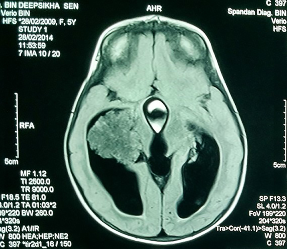
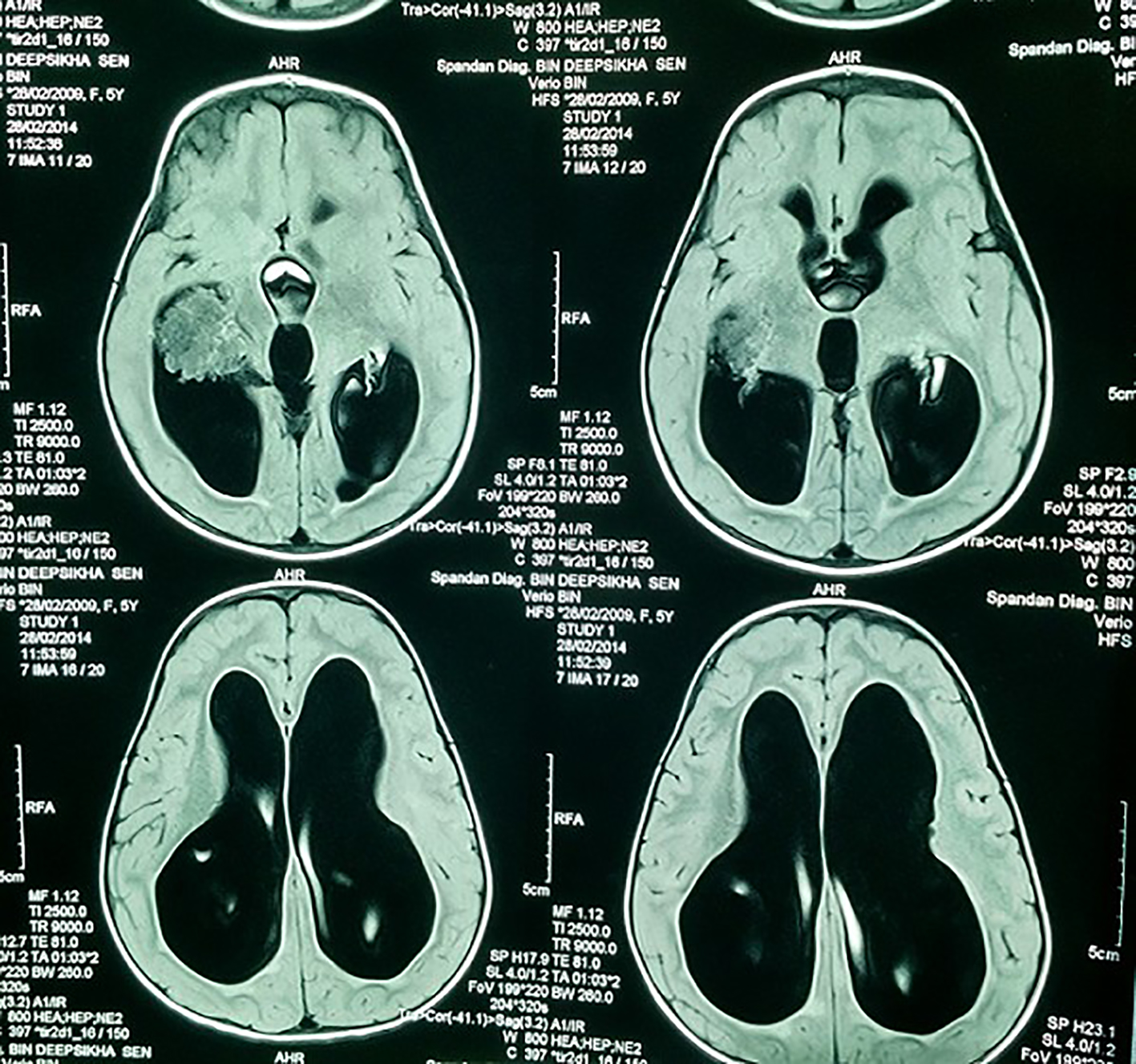
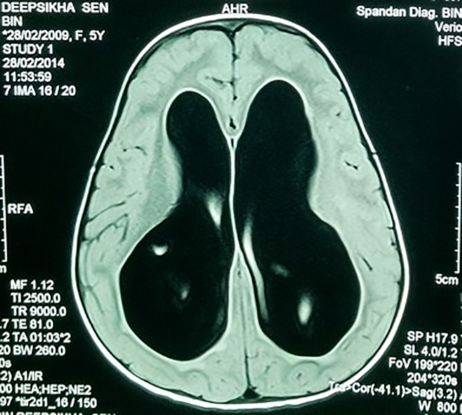
Figure 1. MRI showing large bilateral hydrocephalus.
Figure 2.
Figure 2. MRI showing the hypointense lobulated mass in the lateral ventricle.
Figure 3.
Figure 3. MRI showing lobulated mass and the dilated ventricles.
Figure 4.
Figure 4. Histopathological image showing fibrovascular fronds lined by cuboidal to low columnar cells with extreme area of calcification and focal areas of chondroid and osseous metaplasia in the stroma.
Figure 5.
Figure 5. Immunopositivity for cytokeratin (CK7).
Figure 6.
Figure 6. Strong cytoplasmic immunopositivity for S-100.