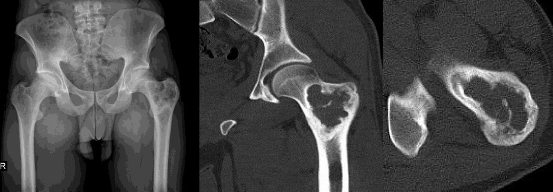
Figure 1. Preoperative X-ray and CT views of the mass. CT: computed tomography.
| World Journal of Oncology, ISSN 1920-4531 print, 1920-454X online, Open Access |
| Article copyright, the authors; Journal compilation copyright, World J Oncol and Elmer Press Inc |
| Journal website http://www.wjon.org |
Case Report
Volume 8, Number 6, December 2017, pages 196-198
Epithelioid Angiosarcoma in Femur: A Case Presentation
Figures