World Journal of Oncology, ISSN 1920-4531 print, 1920-454X online, Open Access
Article copyright, the authors; Journal compilation copyright, World J Oncol and Elmer Press Inc
Journal website http://www.wjon.org

Case Report

Volume 9, Number 4, August 2018, pages 115-118


The First Report of Small Cell Cancer of the Uvula Presenting With Ectopic Adrenocorticotropic Hormone Syndrome

Figures

Figure 1.
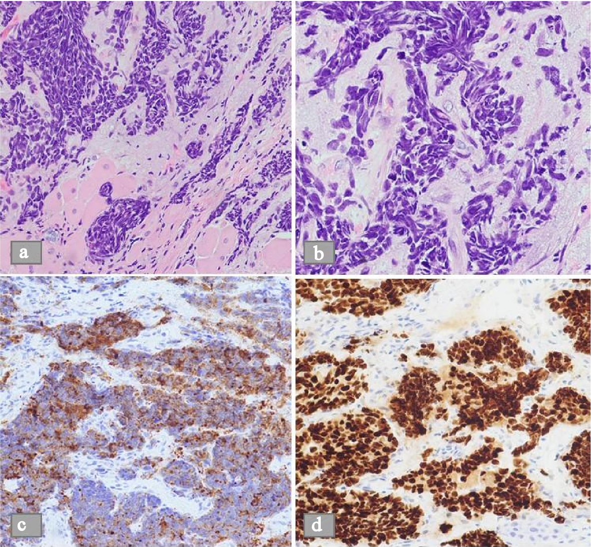
Figure 1. Microscopic view of a histological biopsy of the uvula. (a) H&E stain shows sheets of spindle shaped cells in nests on low power microscopic view (b) H&E stain shows the dense nuclei with inconspicuous nucleoli and scant cytoplasm on high power microscopic view. (c) Demonstrable positivity to synaptophysin stain. (d) Similar positivity to TTF1 stain.
Figure 2.
Figure 2. Magnetic resonance imaging (MRI) of the brain showed heterogeneous, mildly enhancing, T2 bright mass along the left fossa of Rosenmuller, compatible with malignancy (arrows).
Figure 3.
Figure 3. CT scan of the abdomen showed nodular cirrhotic liver with innumerable metastatic deposits within the liver measuring up to 3.2 cm.