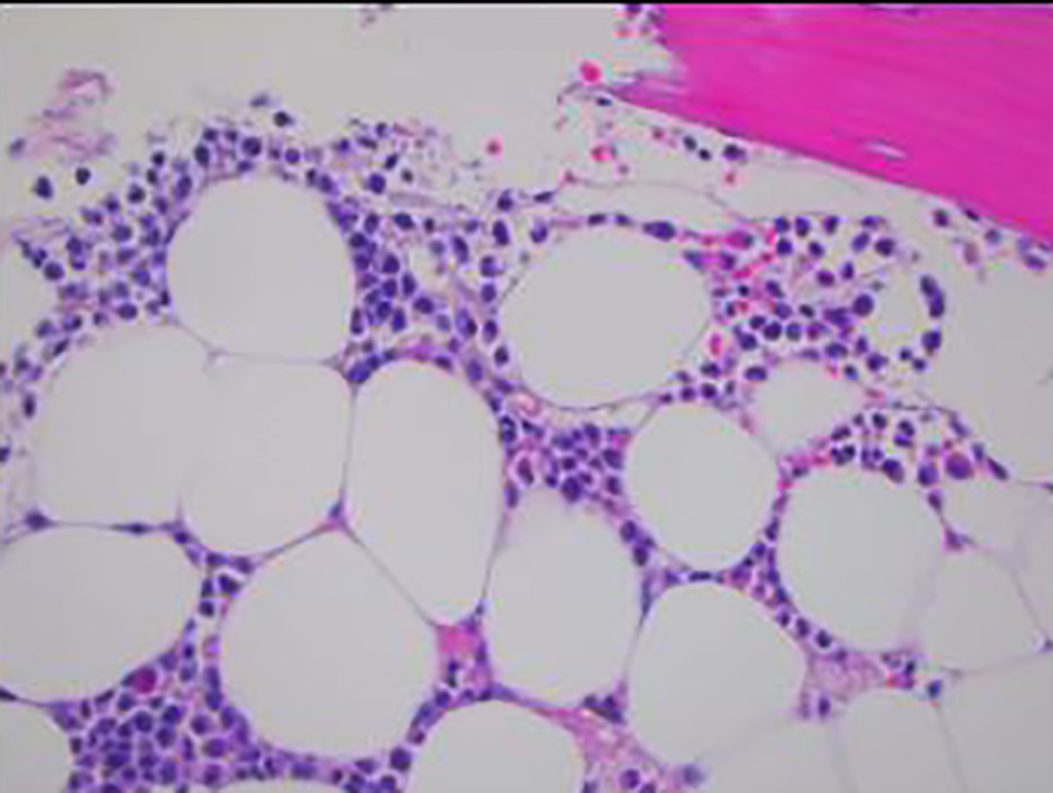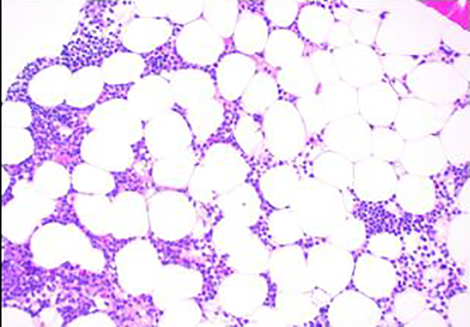
Figure 1. Bone marrow biopsy showing hypocellular marrow for age (× 100).
| World Journal of Oncology, ISSN 1920-4531 print, 1920-454X online, Open Access |
| Article copyright, the authors; Journal compilation copyright, World J Oncol and Elmer Press Inc |
| Journal website http://www.wjon.org |
Case Report
Volume 10, Number 3, June 2019, pages 153-156
Acute Myeloid Leukemia Acquiring Promyelocytic Leukemia-Retinoic Acid Receptor Alpha at Relapse
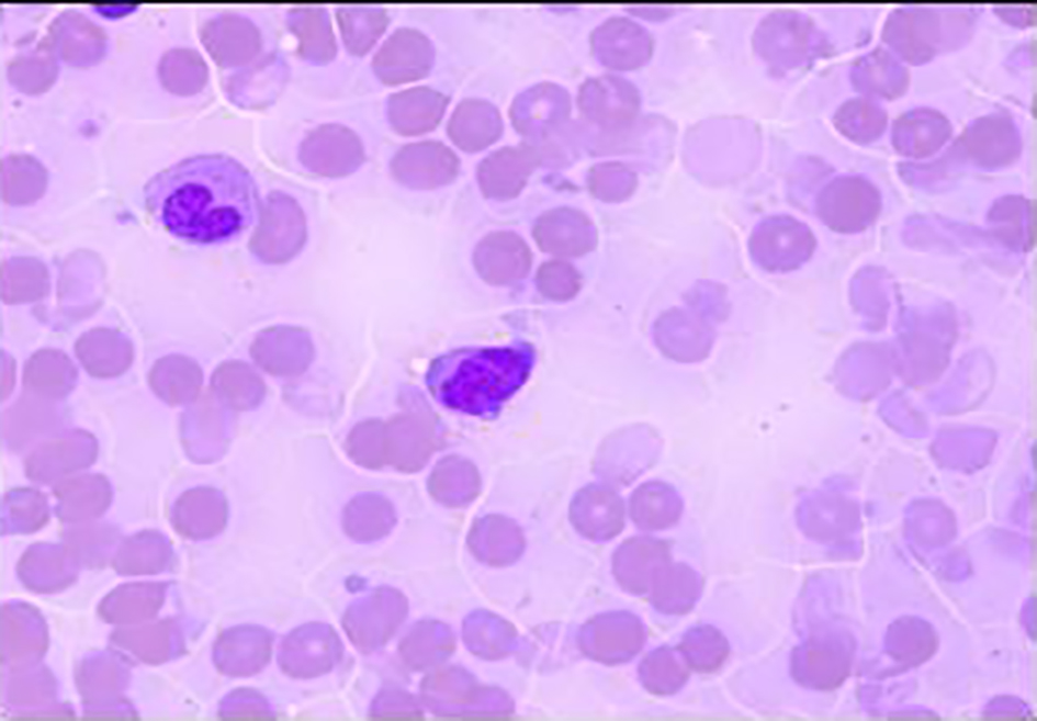
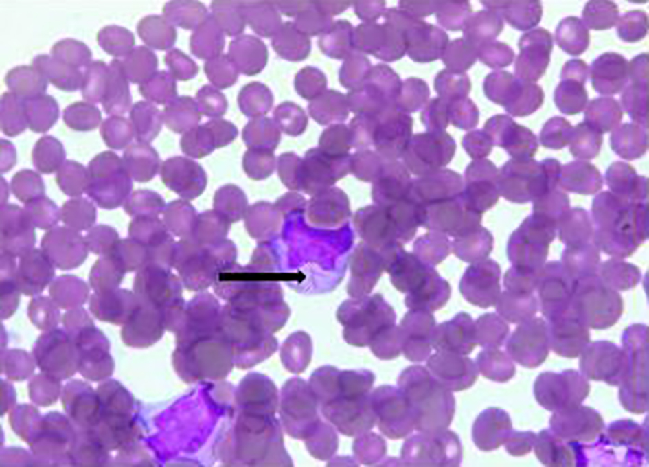
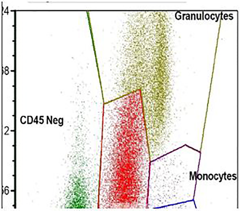
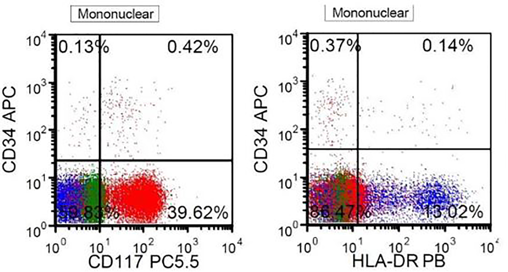
Figures