Figures
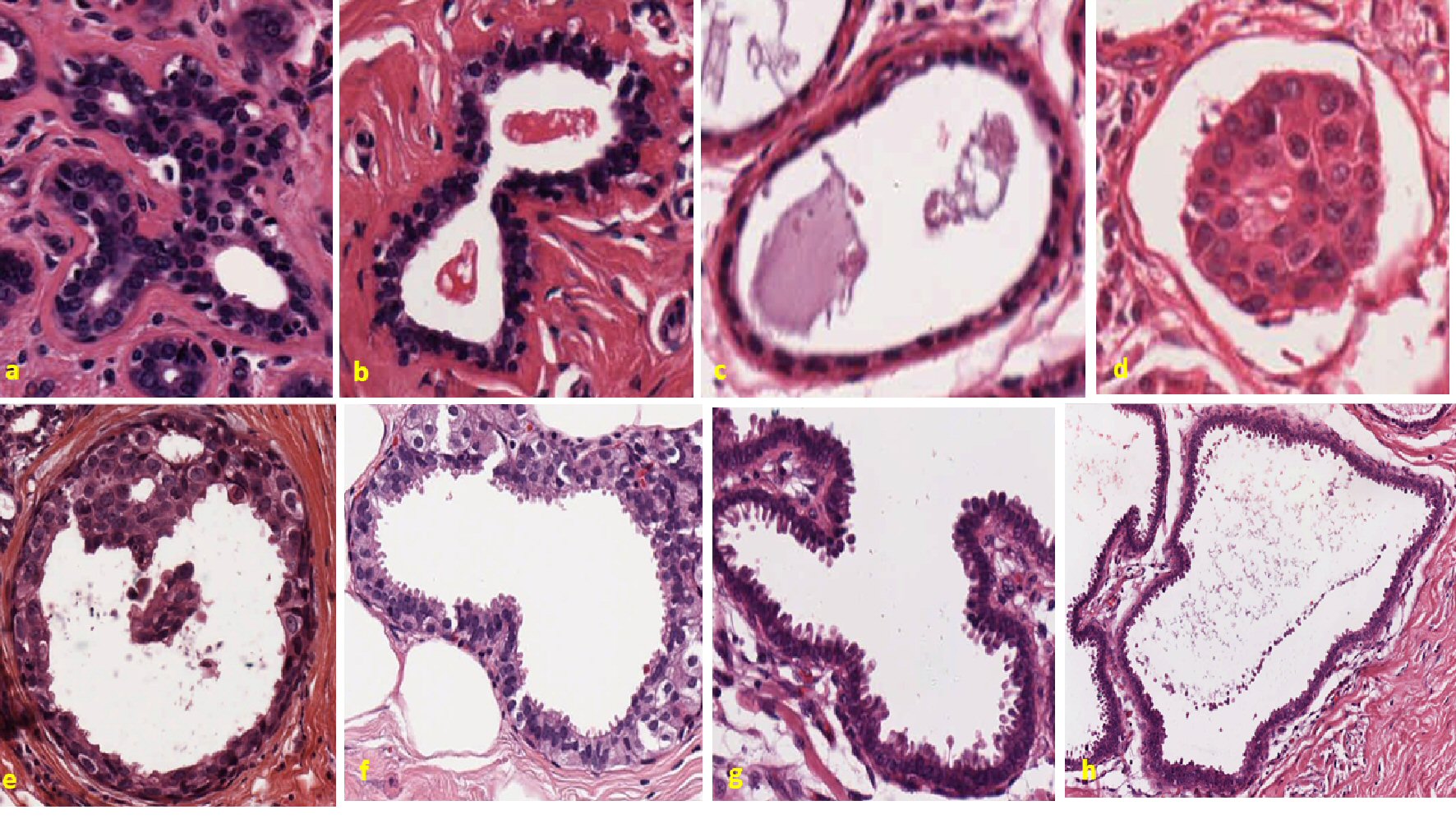
Figure 1. Pleomorphic changes in the cell membrane and egression of excrescents in benign and invasive breast tumors. Representative figure showing control tumors in the upper panel. Figure (a) represents a case of breast giant fibroadenoma. The slide shows well-circumscribed fibroepithelial growth, largely pericanalicular. Uniformly stained nucleus and cytoplasm are seen. The stroma is rather poor in cells and hyaline instead of myxomatous. Next figure (b) also represents another case of breast fibroadenoma. Well-organized cytoplasm and nucleus can be seen. Continuous cellular boundary and no apparent breakage are visible in the tissue. The stroma is fibrous and does not appear sarcomatous. Epithelial cells are not anaplastic. Figure (c) represents a case of breast fibromatosis. The cells are uniformly stained and no randomness in the arrangement of cytoplasm and nucleus is seen. The picture is one of relatively acellular fibrous tissue with a little fat and even less parenchyma, and no cancerous cell is observed in the slide. The last figure in the upper panel figure (d) represents Paget’s disease of breast. There are no apparent dysmorphic changes in membrane and nuclear organization. Lower panel shows different cases of highly metastatic breast cancers. First figure in lower panel figure (e) shows intraductal carcinoma of breast. Highly random cell organization with different shapes and morphology of nuclei are seen. Cells seem like breaking from the basement membrane and some clusters of cells are seen floating inside the lumen. Next figure in the lower panel figure (f) is a different case of intraductal carcinoma of breast. Asymmetric nucleus with formation of vesicular structure is seen unevenly throughout the margin. Breach in the basement membrane is clearly visible; also, some individual cells seem floating inside the lumen. Figure (g) is a case of invasive lobular carcinoma breast. Convoluted membrane structure with lots of villi-shaped cells are projecting outward is clearly seen in this slide. The cells and especially the nuclei are considerably larger than normal or than those of benign proliferative lesion. Cell shapes are distorted and start of stromal infiltration is clearly visible. The case in the lower panel figure (h) is the same case of invasive lobular carcinoma but different region. Most of the features are same as those of previous slide. The cells are further distorted by the start of disintegration and stromal infiltration, and it seems like invasion appears to have progressed. Clusters of cells seem like shedding in pattern and going away from membrane.
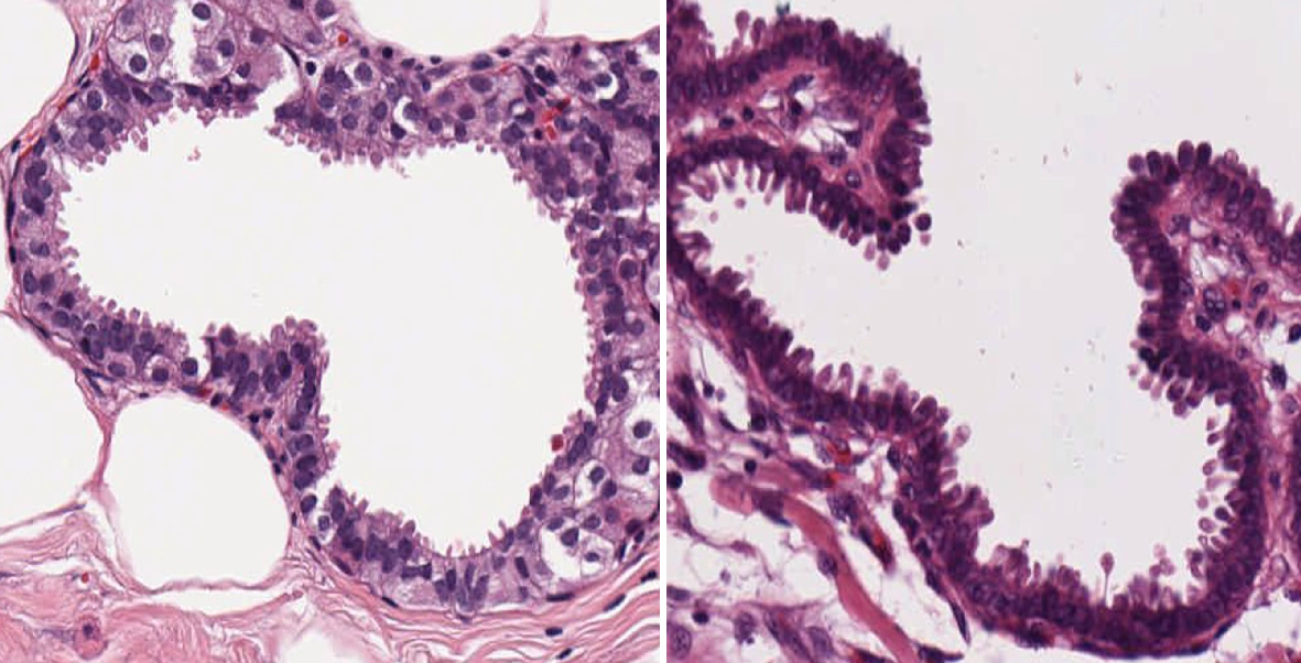
Figure 2. Random projection of vesicles surrounding the membrane. These figures show invasive intraductal carcinoma and invasive lobular carcinoma breast with lots of protrusion surrounding the boundary and with large number of excrescences or finger-like projections projecting towards the lumen randomly throughout the membrane.
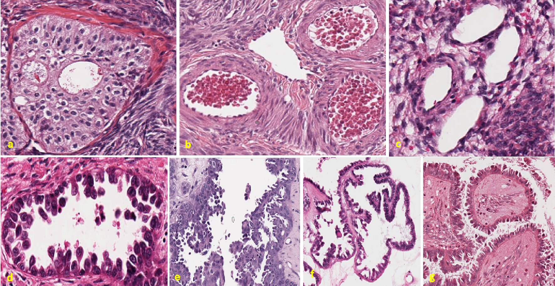
Figure 3. Ovarian tumors showing random projection of vesicles throughout the membrane. Representative figure in the upper panel showing control ovarian cases. The first figure in the upper panel figure (a) is a case of benign Brenner tumor of ovary. There is no apparent protrusion or degradation of the membrane. Cells are uniformly stained throughout the slide. Next figure (b) is a case of polycystic ovarian disease. Well-organized tissue slides with no malignant cells are visible in the slide. Last figure in the upper panel figure (c) represents a case of benign thecoma ovary. Evenly distributed cytoplasm and nucleus are seen, no membrane disruption is observed. Lower panel shows all metastatic cases of ovarian tumor. Figure (d) represents a case of endometrioid carcinoma ovary. Ball and stick-like structure are consistent throughout the slide and cells can be seen in different phase of their division. Cells seem like breaking and floating inside the lumen. Next figure (e) represents high-grade carcinoma ovary with diagnosis of serous, clear cell and endometrioid tumor. Highly random tissue organization and breakage of basement are clearly visible in this slide. Also, cells are floating in cluster or a single cell inside the lumen. Next figure (f) represents a case of serous carcinoma ovary. The arrangement of basement membrane is convoluted and villi-like cell projection can be observed throughout the slide. Last figure in the lower panel figure (g) also represents a case of high-grade serous carcinoma ovary. There is unevenly finger-like projection in the tumor membrane outside of the lumen. Random projections of vesicles surrounding the membrane in different structure and shape are seen.
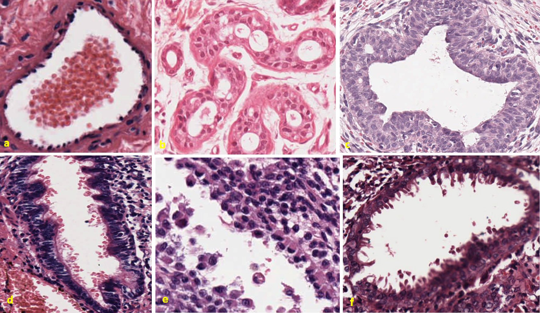
Figure 4. Different types of cancer have same morphological features of sprouting throughout the membrane. Figure (a) shows control lung hamartomatosis. Well-organized boundary throughout the slides is clearly visible. No randomness among the cells is observed. Next figure (b) represents a case of benign Bowen’s disease of skin. Uniformly stained cells are observed all over the slide. No sign of breakage of basement membrane is observed. Next figure (c) represents a cellular giant fibroadenoma (benign cystosarcoma phylloides left breast) with organized cellular structure of cytoplasm and nucleus, respectively. In lower panel, the first figure (d) shows giant bone cell tumor, metastatic to the lung. Cells are highly disorganized and sprouting out from the membrane towards lumen. Cells are seen in different phase of division. Some individual cells are floating in the lumen. Next figure (e) shows metastatic malignant melanoma ovary. Highly disorganized cells with euchromatin and heterochromatin nuclei. Some cells have apical nuclei, while other do not. Cells are also breaking from the membrane and floating in clusters. Last figure (f) shows cystosarcoma phylloides of the breast. Highly pleomorphic nucleus and randomly scattered vesicular structures are visualized floating inside the lumen. This tumor has random and different phasic nuclei, irregularly dispersed cells with processes and vesicles are extending towards lumen.

Figure 5. Metastatic tumors have convoluted and crooked margins than the control tumors, which have well-formed defined cellular margins. Representative figures in the upper panels showing controls. The first figure is showing control original H&E staining of lung hamartomatosis, the next one is of fibroadenoma breast, and the next two images is of margins of the same control tumor represented with ImageJ perimeter measurement. White pseudocolored margins are well defined with no breakage or distortions throughout the perimeter. In the lower panel, the first two images show original H&E staining of giant cell metastatic bone tumor to the lung, and invasive lobular carcinoma of the breast; the next two figures are the representation of margins with images of same tumors. Margins of metastatic tumors are highly crooked, distorted and not well defined. Orange arrows in the figures indicates waviness of the membranes of metastatic tumor in contrast with control tumor. H&E: hematoxylin and eosin.
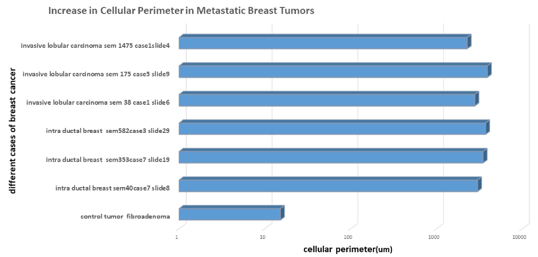
Figure 6. Quantitative measurement of increase in membrane perimeter in invasive cancer. There is distinctive increase in membrane perimeter in invasive breast tumor measured with the ImageJ tools compared with benign breast case. Increase in perimeter is shown in blue bar graph with µm unit. There is nearly 100-fold increase in length of perimeter in invasive breast tumor.
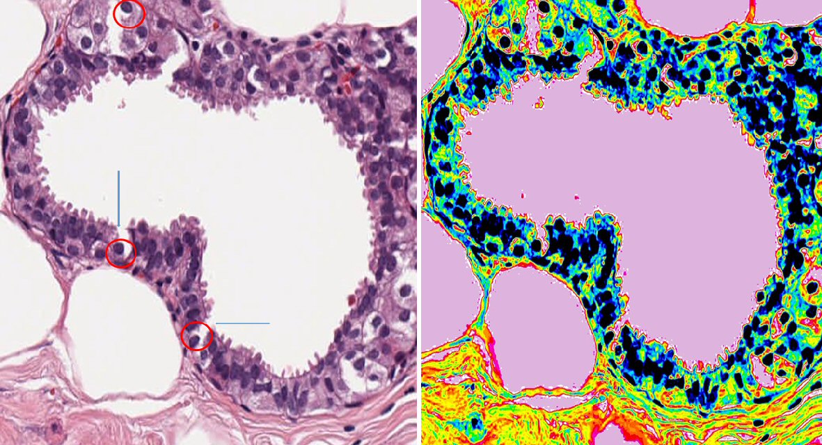
Figure 7. Breaching of basement membrane is more apparent in the tumor where there are lot of excrescences sprouting along the membrane in invasive breast cancer. This figure shows the microscopic image of invasive intraductal carcinoma breast. Figure on the left is the original image of tumor slide with × 20 magnification. Figure on the right-hand side is the pseudocolored image of the same slide made using ImageJ for clear demonstration purpose. This has random breakage of basement membrane. There are numerous dysplastic cellular arrangements through the lumen and marked nuclear polymorphism. Breakage of basement membrane is found also uncovering the nucleus, leaving it naked (represented with red circle) and with blue arrow. There are some floating vesicles seen in the lumen. The next panel shows pseudocolored image of the same slide, where floating vesicles is clearly visible.
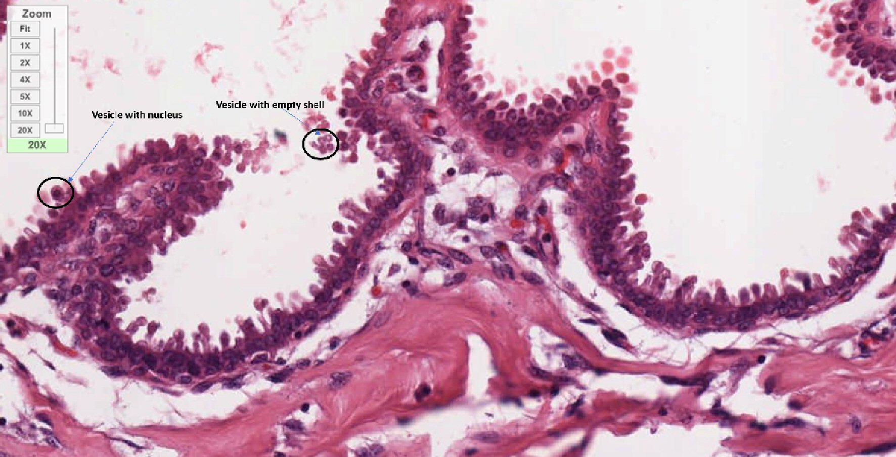
Figure 8. Distinct mushroom-like projections with oval head and cylindrical stalk coming out from membrane. This figure is a × 20 magnified image of invasive lobular carcinoma of breast. Finger-like vesicular projections are seen coming out from the membrane. All these vesicles are different in length but have almost uniform morphology. Some of them are attached with membrane and others are detaching from the membrane and migrating towards the lumen in cluster. Some of these vesicles are just empty and some of them carrying nucleus as marked with black circle and blue arrow.
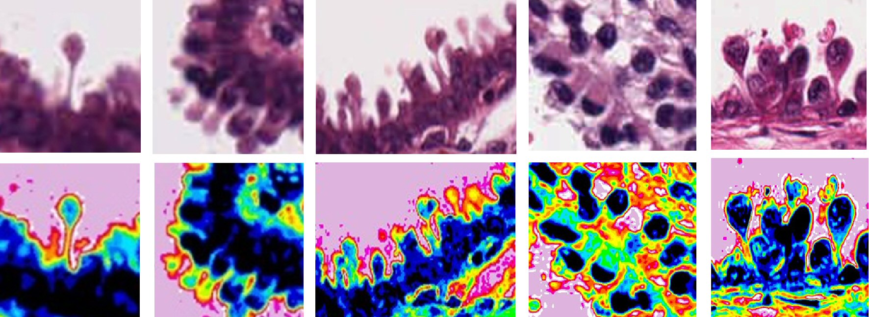
Figure 9. Different shapes and morphological structure of budding excrescents from different invasive cancers (First three panels, invasive lobular carcinoma of breast; malignant melanoma; ovarian endometrioid tumor). This figure shows different forms of budding vesicles in invasive cancers. Upper panel shows all magnified original image of excrescents, while lower panel shows pseudocolored image of the same tissue done with ImageJ 16-color options corresponding image with upper panel. All these projections have common rounded or oval-shaped head with cylindrical stalk. Some of them are well formed, while others are in the process of formation. Few of these structures are with nuclei, while some are without nuclei. The morphology of these projections is quite random throughout the perimeter.