World Journal of Oncology, ISSN 1920-4531 print, 1920-454X online, Open Access
Article copyright, the authors; Journal compilation copyright, World J Oncol and Elmer Press Inc
Journal website http://www.wjon.org

Case Report

Volume 11, Number 3, June 2020, pages 116-121


Small Bowel Gastrointestinal Stromal Tumor as a Gateway for Streptococcus anginosus Causing Multiple Liver Abscesses

Figures

Figure 1.
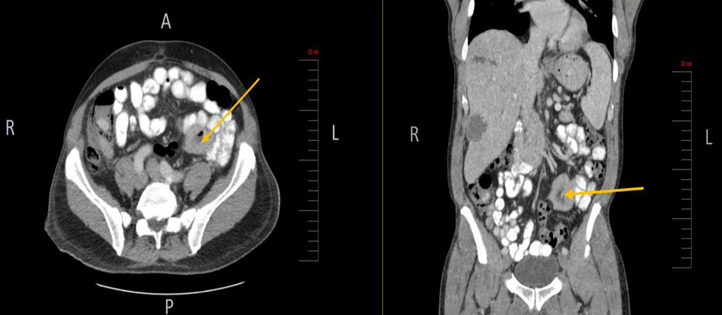
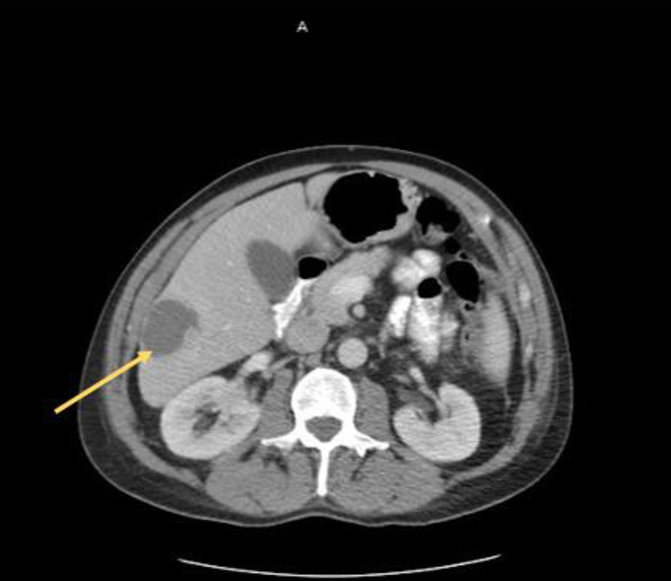
Figure 1. Liver lesion (arrow) identified on axial CT scan of the abdomen and pelvis. CT: computed tomography.
Figure 2.
Figure 2. Small bowel mesenteric mass (arrow) identified on CT scan of the abdomen and pelvis (axial and coronal views, respectively). CT: computed tomography.
Figure 3.
Figure 3. Small bowel mass at the mesenteric root, status post biopsy.
Figure 4.
Figure 4. Biopsy of small bowel GIST shows proliferation of bland appearing spindle cells with eosinophilic fibrillar cytoplasm arranging in short fascicular pattern, with minimal pleomorphism and low mitotic index (< 5/50 HPF) (a, × 10 objective magnification). The spindle cells are immuno-positive for CD117 (c-KIT) (b). GIST: gastrointestinal stromal tumor.