World Journal of Oncology, ISSN 1920-4531 print, 1920-454X online, Open Access
Article copyright, the authors; Journal compilation copyright, World J Oncol and Elmer Press Inc
Journal website http://www.wjon.org

Case Report

Volume 11, Number 3, June 2020, pages 122-125


Dural Marginal Zone Lymphoma in a Patient With a Hepatitis C Virus Infection

Figures

Figure 1.
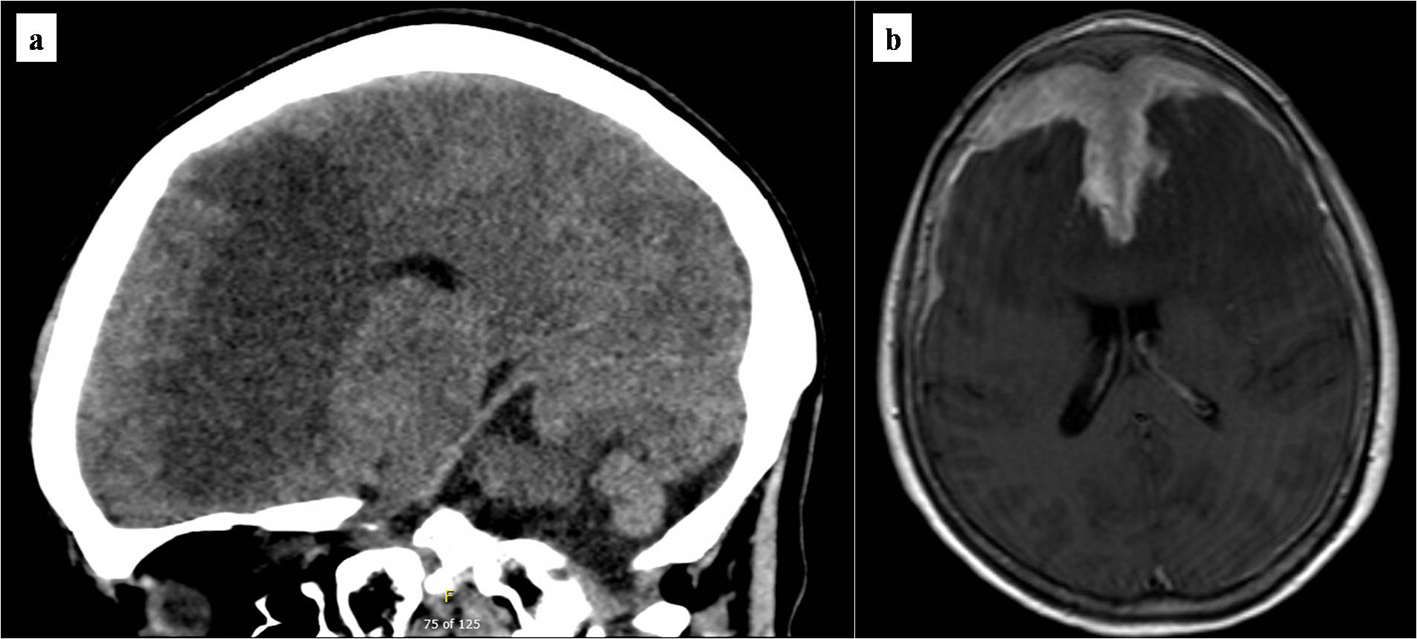
Figure 1. Brain imaging at presentation. (a) Initial non-contrasted computed tomography scan of the brain. Marked bifrontal white matter hypodensity extends across the corpus callosum with mass effect on the frontal horns of the lateral ventricles. In addition, there is a large poorly marginated region of hyperdensity along the anterior falx and anterior cranial vault which appears extra-axial. (b) T1/2-weighted brain MRI. Bifrontal extra-axial mass with extensive dural tail with invasion into both frontal lobes and extensive edema extending across the corpus callosum. MRI: magnetic resonance imaging.
Figure 2.
Figure 2. Histopathology of dural biopsy. Microscopic examination shows sheets of small lymphoid cells with clumped chromatin and monocytoid morphology (hematoxylin and eosin, × 400) (a). The lymphoid population is composed predominantly of CD20-positive B cells (b) which aberrantly express CD43 (c) (CD20 and CD43 IHC, × 400). Frequent interspersed small CD3-positive T cells are also present (CD3 IHC, × 400) (d). IHC: immunohistochemistry.