Figures
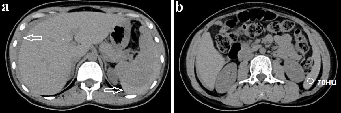
Figure 1. Axial abdominal CT pre-contrast images. (a) Free intraperitoneal fluid (arrow) was detected. (b) The fluid surrounding the spleen was hyperdense, measuring 70 Hounsfield unit (HU), suggesting blood within the peritoneal cavity. CT: computed tomography.
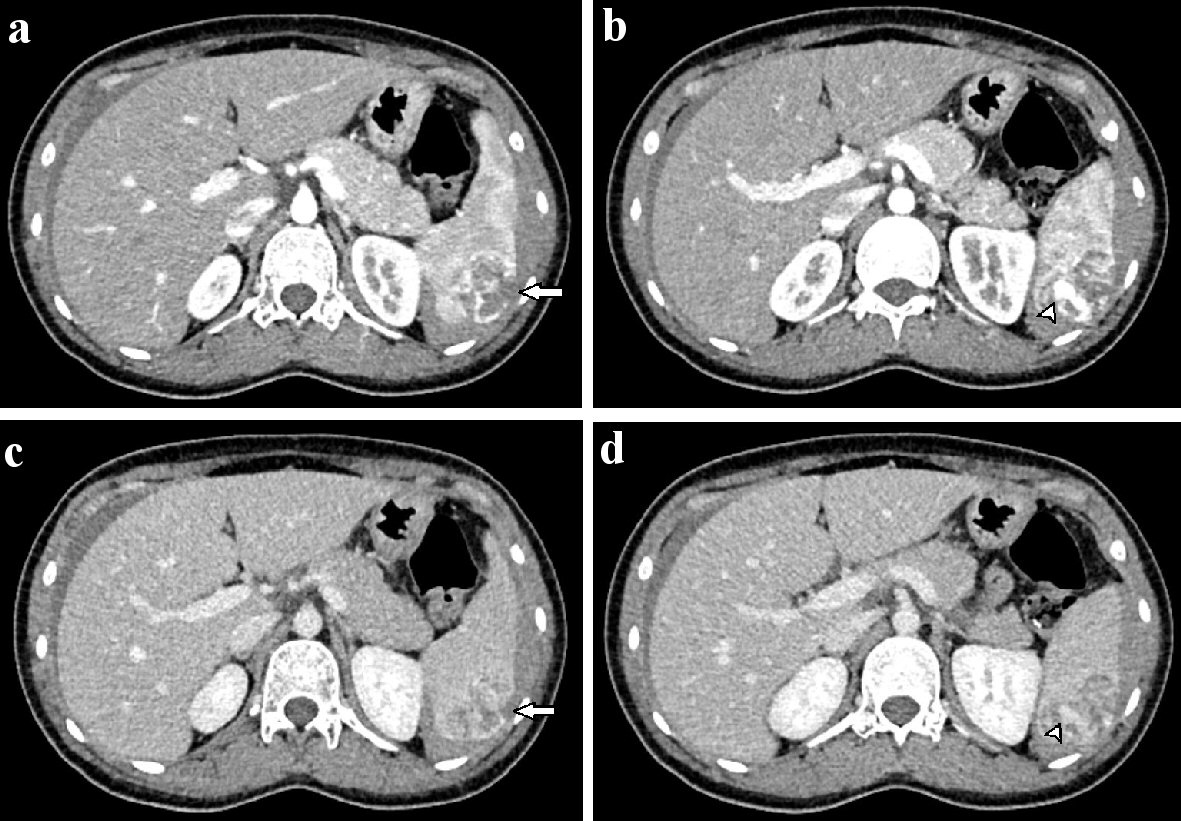
Figure 2. Axial abdominal CT in the arterial (a, b) and venous phases (c, d). A 3 × 4 cm mass with ill-defined borders was observed in the spleen (a). This mass showed heterogeneous enhancement and hypervascularity (a, b). The mass enhanced heterogeneously on the venous phase (c). A tumor laceration was observed on the splenic capsule (a, c), resulting in intratumor pseudoaneurysm (b, d; arrowhead). CT: computed tomography.
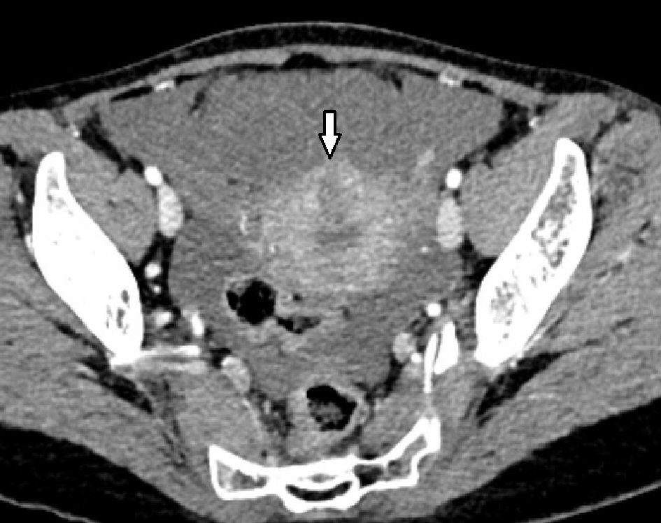
Figure 3. A hypoenhancing mass was observed on the uterine wall.
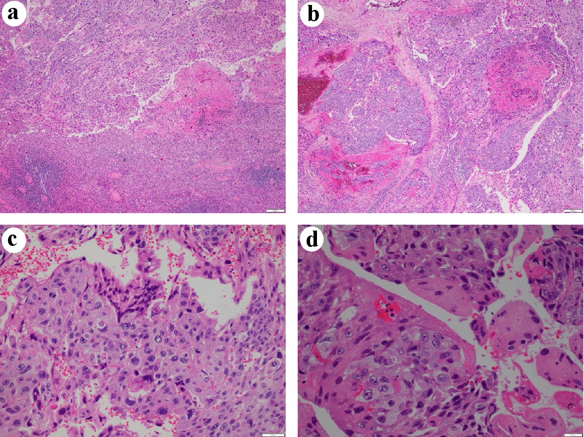
Figure 4. Hematoxylin and eosin (H&E) staining. (a) The tumor was solid, with large dyscohesive and pleomorphic cells (× 40). (b) Hemorrhagic and necrotic areas were observed (× 40). (c) Two types of identified cells: oval and giant cells, similar to cytotrophoblastic cells and syncytiotrophoblastic cells, respectively (× 200). (d) Cytologic atypia and mitotic figures (× 400).
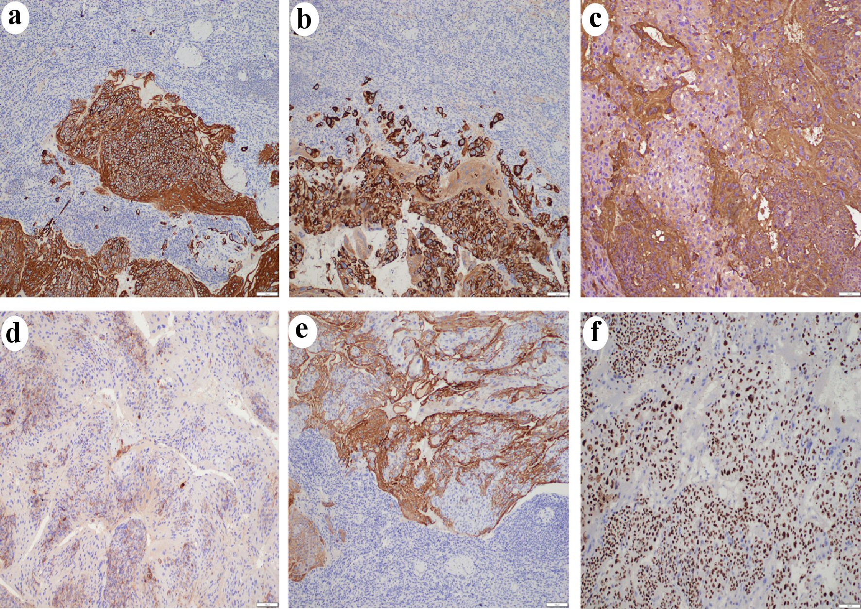
Figure 5. Immunohistochemistry results (× 100) showed tumor cells positive for CK (a), CK7 (b), HCG (c), PLAP (d), and inhibin (e). A high Ki-67 labeling index (> 90%) was observed (f). CK: cytokeratins; CK7: cytokeratin 7; HCG: human chorionic gonadotropin; PLAP: placental alkaline phosphatase.