World Journal of Oncology, ISSN 1920-4531 print, 1920-454X online, Open Access
Article copyright, the authors; Journal compilation copyright, World J Oncol and Elmer Press Inc
Journal website https://www.wjon.org

Case Report

Volume 12, Number 1, February 2021, pages 34-38


Clear Cell Carcinoma of the Cervix With OHVIRA Syndrome: A Rare Case Report

Figures

Figure 1.
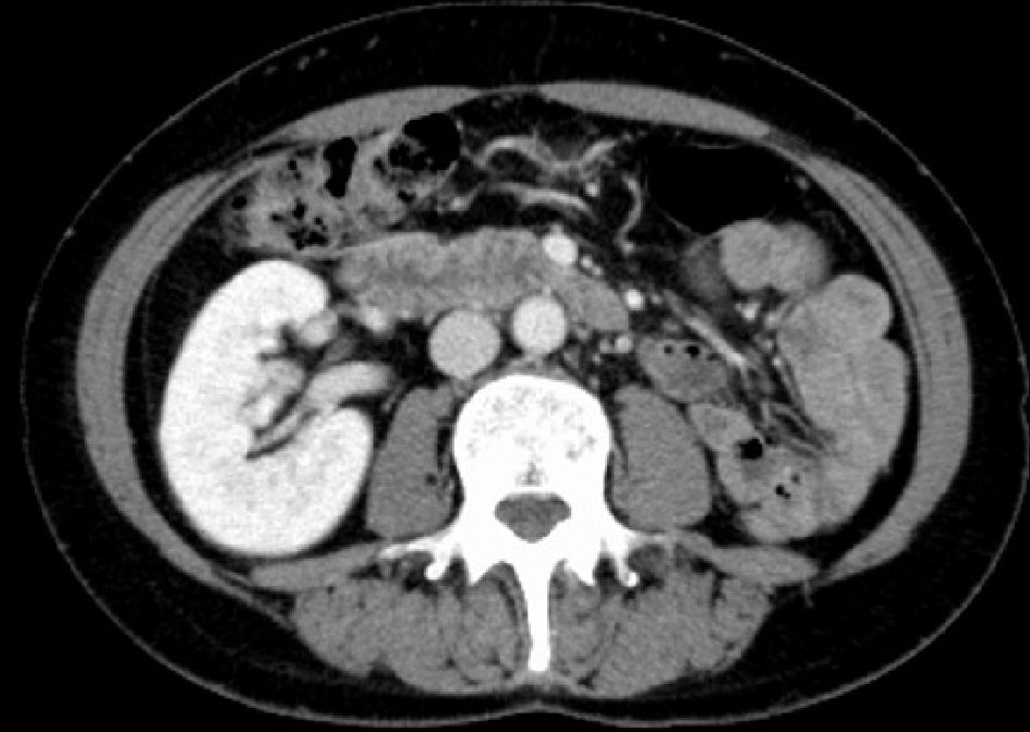
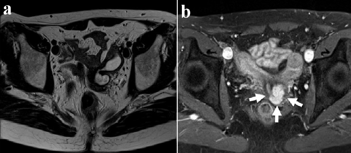
Figure 1. Axial view of pelvic magnetic resonance imaging (MRI). T2-weighted MR image shows the left obstructed hemivagina (a). T2-weighted postcontrast MR image shows uterine didelphys and a tumor measuring 1.0 × 2.0 cm on the left side of the uterine cervix (white arrows) (b).
Figure 2.
Figure 2. CT scan shows the absence of the left kidney. CT: computed tomography.
Figure 3.
Figure 3. Surgical specimen shows a cervical tumor with uterus didelphys and her left ectopic ureteral orifice in the left obstructed hemivagina. A tumor is detected on the left side of the uterine cervix (white arrows). The surgical sonde was passed through the left ectopic ureter.
Figure 4.
Figure 4. Histopathological findings of cervical clear cell carcinoma. At low-power view, the tumor shows papillary growth (a) and tubulo-cystic growth pattern (b) (hematoxylin and eosin stain (H&E), × 40). At high-power view, tumor cells have enlarged nuclei with prominent nucleoli and clear cytoplasm (c) (hematoxylin and eosin stain (H&E), × 400). Immunohistochemical staining show the tumor cells are positive for napsin A, × 200 (d); and HNF-1β, × 200 (e); whereas p16 block positive pattern (× 200) is not observed in the tumor (f).