Figures
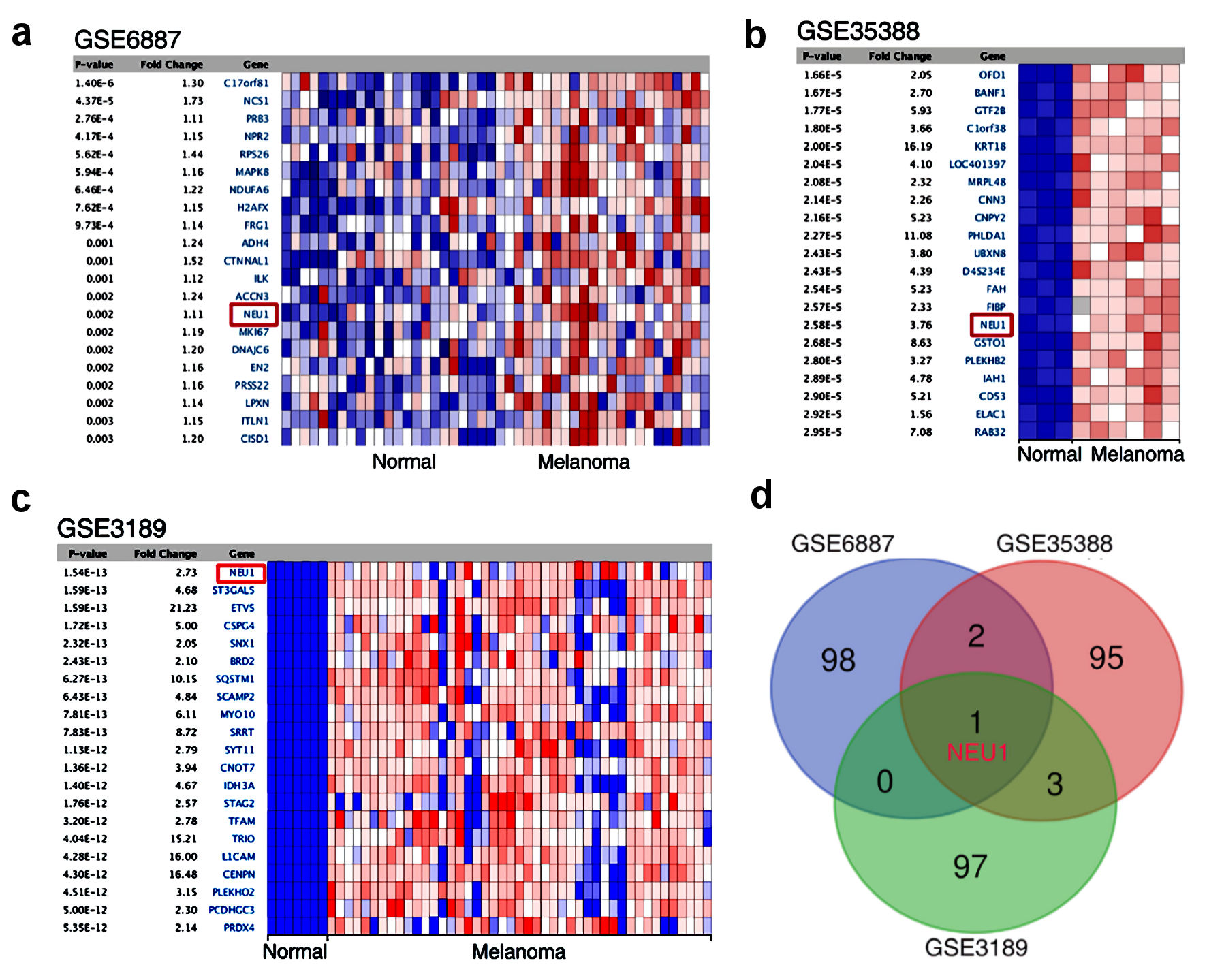
Figure 1. NEU1 was highly expressed in melanoma. (a-c) Heatmap showing genes that are dysregulated in melanoma compared to normal tissues mined from GSE6887, GSE35388 and GSE3189. (d) Venn diagram identified and validated the dysregulated expression genes shared in the three GEO data sets. NEU1: neuraminidase-1.
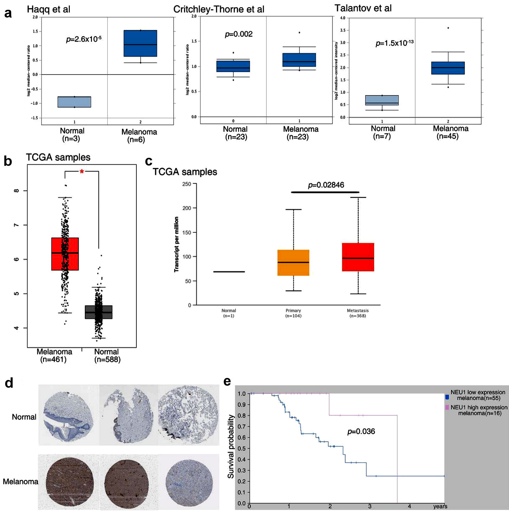
Figure 2. NEU1 expression in melanoma is associated with poor patient survival. (a) Expression data of NEU1 from ONCOMINE database in melanoma samples and controls. (b) Expression data of NEU1 from GEPIA database in melanoma samples. (c) NEU1 expression data in non-cancer controls, primary cancer samples and metastasis melanoma samples from UALCAN database. (d) NEU1 expression data from The Human Protein Atlas database in melanoma patient with or non-cancer controls were measured by immunohistochemical staining. (e) Overall survival rate of melanoma patients in high expression NEU1 group and low expression NEU1 group in The Human Protein Atlas database. NEU1: neuraminidase-1; TCGA: The Cancer Genome Atlas.
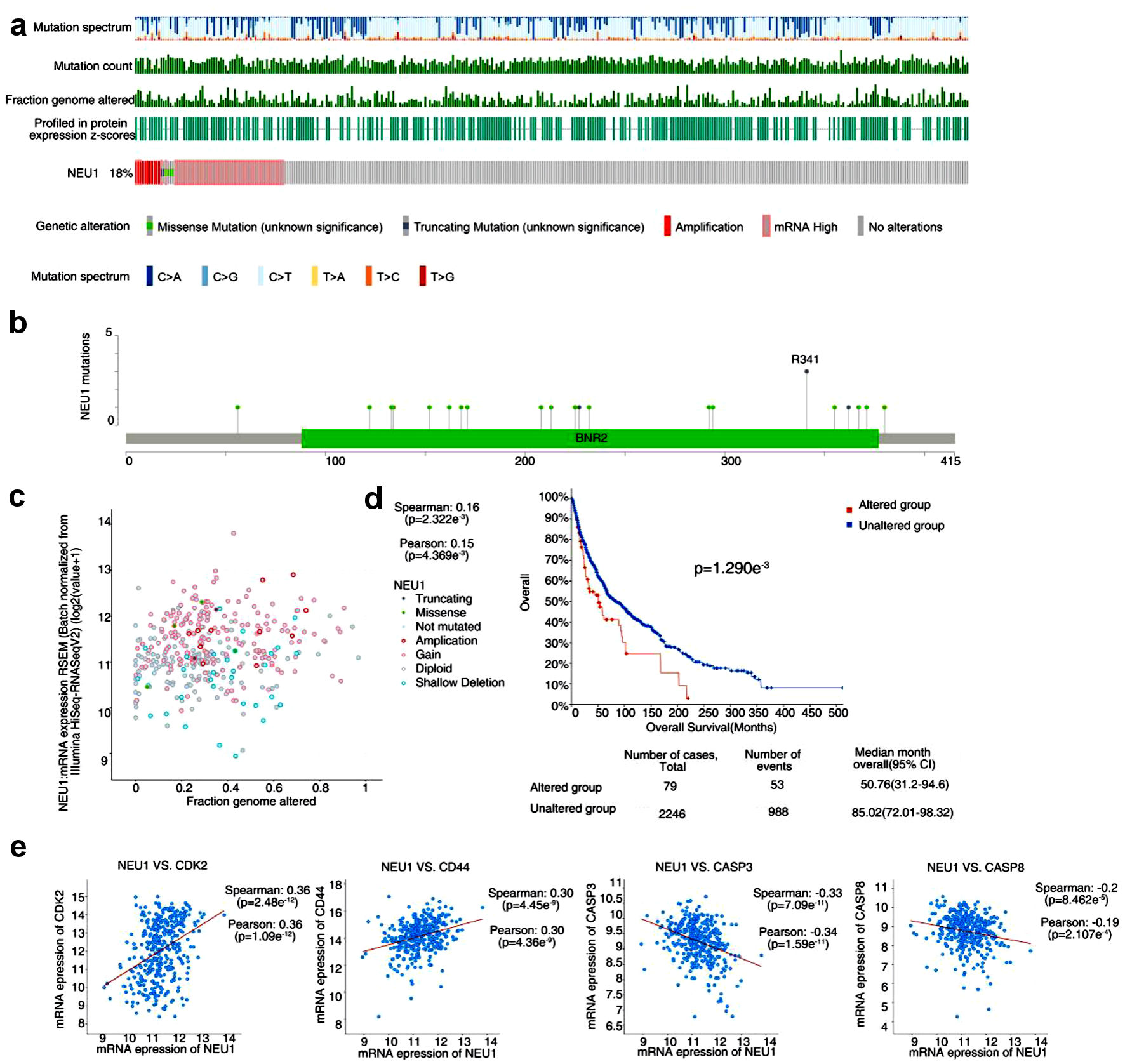
Figure 3. Genetic alteration of NEU1 in melanoma. (a) NEU1 genetic alterations in the melanoma using the cBioPortal tool. (b) The mutation site and mutation frequency of NEU1 were displayed using the cBioPortal tool. (c) Correlation analysis of fraction genome altered of NEU1 with mRNA expression of NEU1 using the cBioPortal tool. (d) Correlation between NEU1 mutation and overall survival in melanoma. (e) Correlation between expression of NEU1 in melanoma and tumor related markers such as CDK2, CD44, CASP3 and CASP8. NEU1: neuraminidase-1.
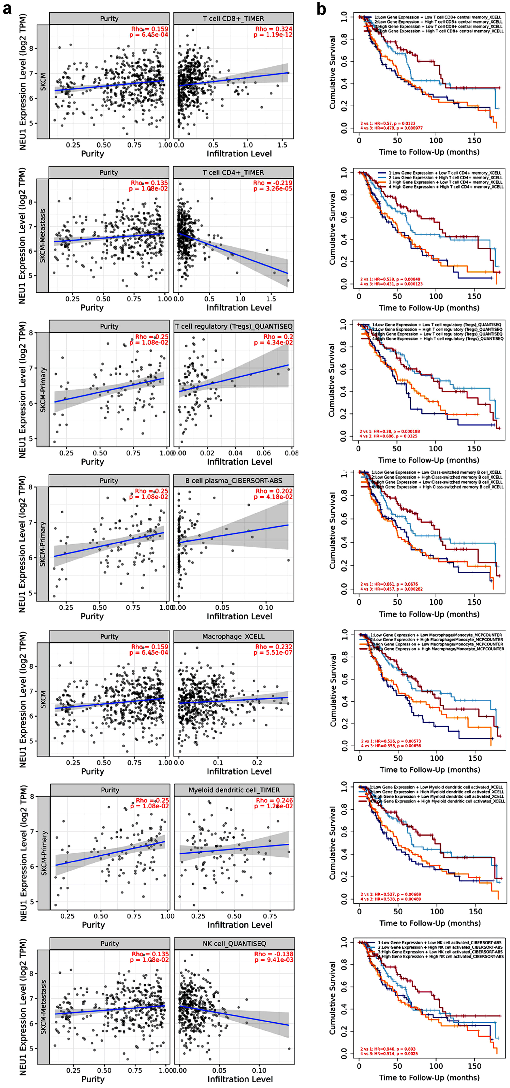
Figure 4. Correlation between NEU1 and immune cell infiltration. (a) The correlation between NEU1 expression and the infiltration of CD4+ T cell, CD8+ T cell, B cell, regulatory T (Treg) cell, macrophage, and natural killer (NK) cell in melanoma was analyzed by TIMER 2.0 tool. (b) The relationship between the immune cell infiltration and the overall survival of melanoma using the TIMER 2.0 tool. NEU1: neuraminidase-1.
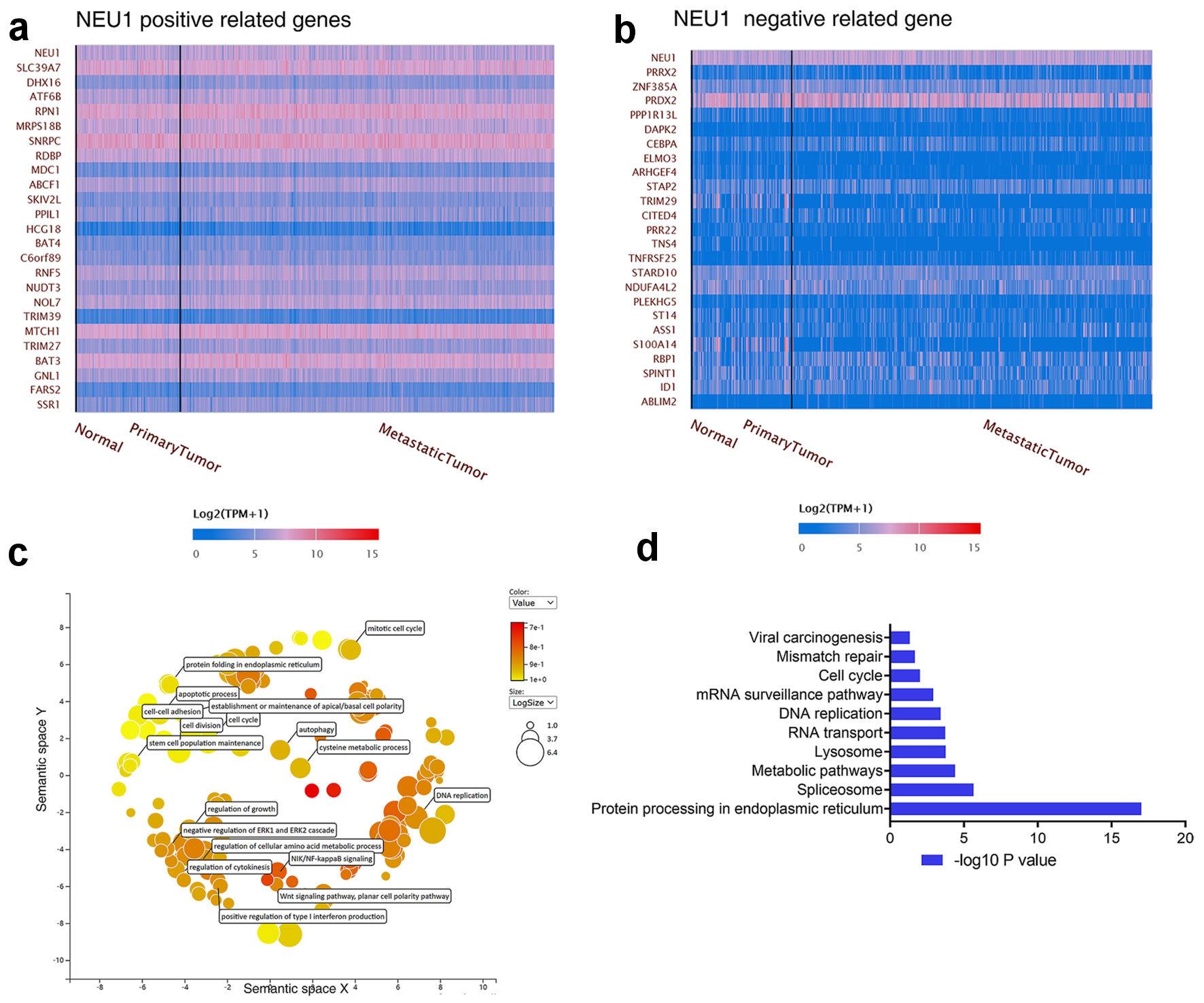
Figure 5. Enrichment analysis of NEU1-related partners. (a, b) The top 20 genes with positive correlation (a) and negative correlation (b) with NEU1 in melanoma were analyzed in the UALCAN database. (c) Functional enrichment histogram of important modules by using REVIGO tools. (d) Pathway enrichment map of NEU1 and NEU1-correlated genes. NEU1: neuraminidase-1.
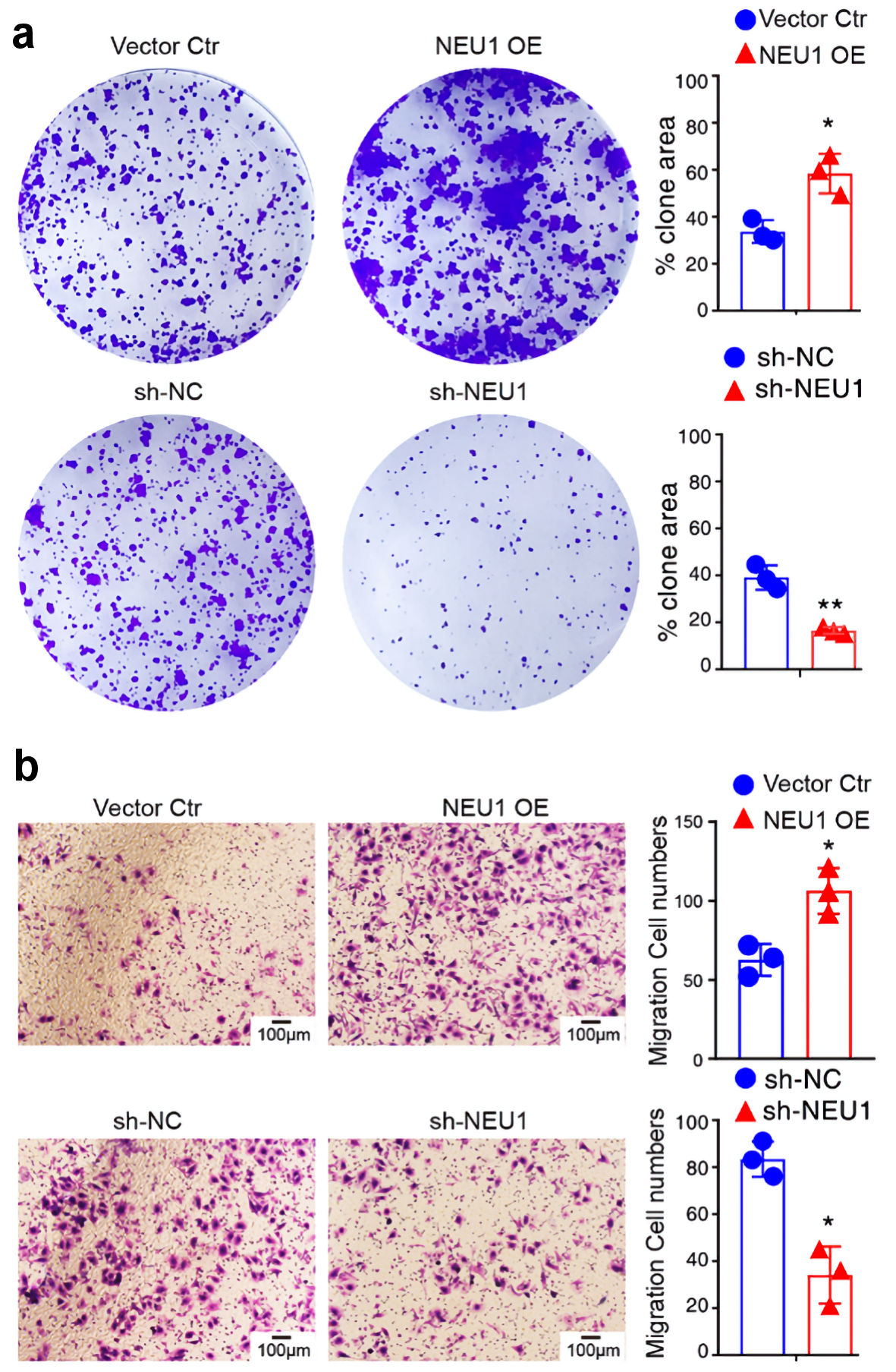
Figure 6. NEU1 promotes melanoma cells growth and migration. (a) Colony formation of A875 cells 2 weeks after transfection with the indicated vector. (b) Transwell assay was used to detect the effect of NEU1 on the migration of A875 cells. NEU1: neuraminidase-1. NEU1: neuraminidase-1.
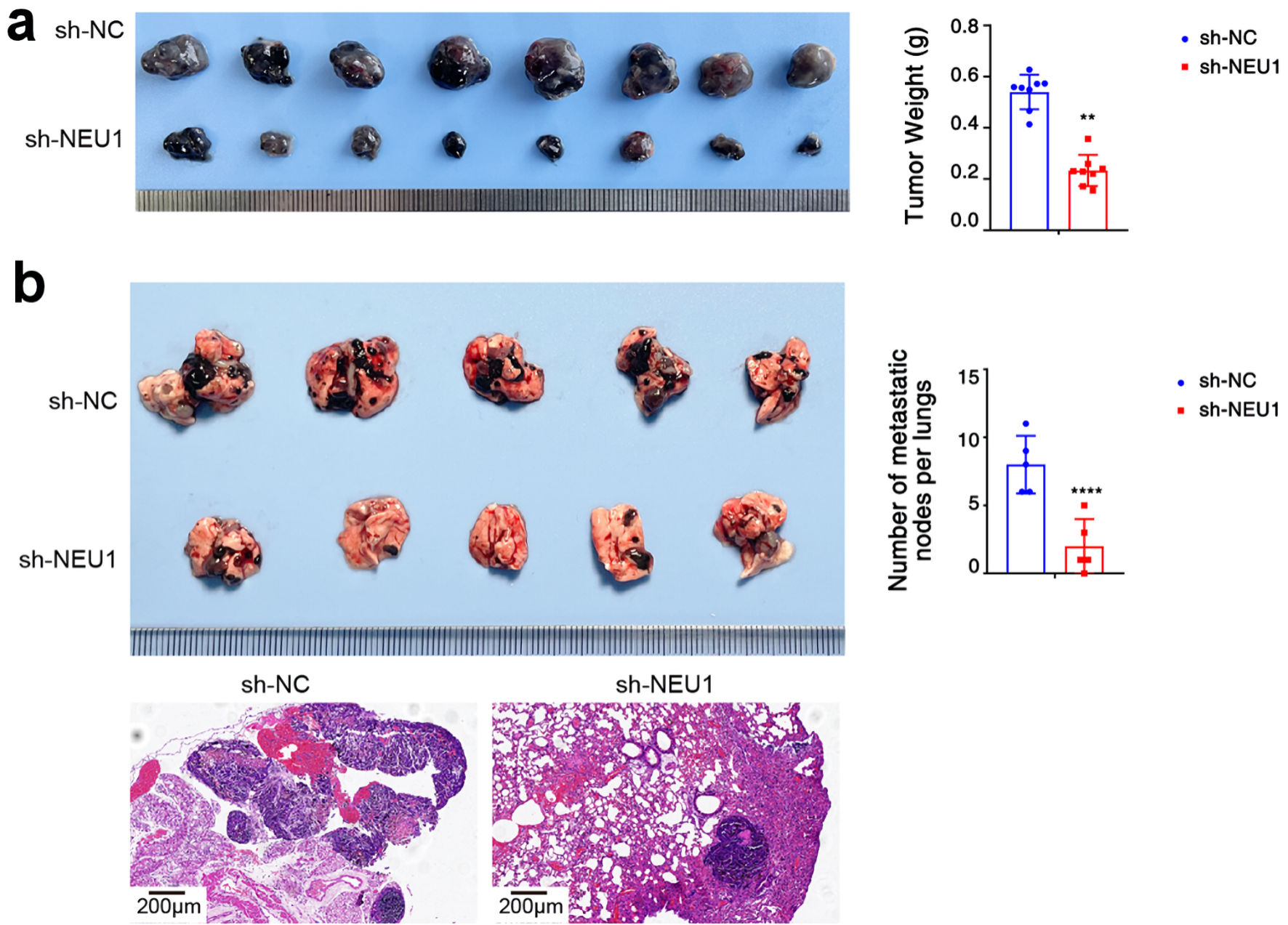
Figure 7. NEU1 promote melanoma cells progression in vivo. (a) Tumor growth of A875 xenografts in nude mice (n = 8 each). Groups 1, 2 mice were injected with NC and sh-NEU1 cells, respectively. (b) After the melanoma cells transfected with sh-NEU1or sh-NC were injected into the tail vein of mice, a representative image of nodules could be seen on the lung surface of mice at 8 weeks (upper panel). Representative images of lung metastasis with H&E staining (lower panel). NEU1: neuraminidase-1. NEU1: neuraminidase-1.