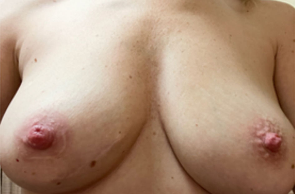
Figure 1. A 40-year-old woman with 2-year history of pain and right nipple enlargement. (a) Macroscopic findings of the nipples. Note the deformity and enlargement of the right nipple compared to the normal left nipple.
| World Journal of Oncology, ISSN 1920-4531 print, 1920-454X online, Open Access |
| Article copyright, the authors; Journal compilation copyright, World J Oncol and Elmer Press Inc |
| Journal website https://www.wjon.org |
Case Report
Volume 13, Number 4, August 2022, pages 235-240
Syringomatous Tumor of the Nipple
Figures
Table
| Case | Author | Age (years) | Sex | Location | Clinical Exam Findings | Size (cm) | Surgery |
|---|---|---|---|---|---|---|---|
| SAN: syringomatous adenoma of the nipple; F: female; M: male; L: left; R: right; NR: not revealed. | |||||||
| 1 | Carter et al, 2004 [5] | 23 | F | Unilateral | Erythematous, tender right nipple nodule with thick, white material extruding from nipple under pressure | 3.5 | Local excision |
| 2 | Yosepovich et al, 2005 [13] | 33 | F | Unilateral | Left nipple mass | NR | Excisional biopsy |
| 3 | Oliva et al, 2008 [14] | 32 | F | Unilateral | Markedly deformed nipple-areolar complex with underlying mass effect | 4 | Central mound excision with a transposition flap closure |
| 4 | Riaz et al, 2008 [15] | 39 | F | Unilateral | Not available | NR | Surgery |
| 5 | Page et al, 2009 [9] | 40 | F | Supernumerary breast | Enlargement of supernumerary breast with milk production postpartum | NR | Punch biopsy |
| 6 | Sharma et al, 2009 [16] | 32 | F | Unilateral | Ill-defined, indurated, non-tender subcutaneous nodule within areolar region | 0.5 | Complete excision |
| 7 | Odashiro et al, 2009 [17] | 44 | F | Unilateral | Irregular nodule on nipple area with edema | 0.75 | Complete excision |
| 8 | Kim et al, 2010 [18] | 48 | F | Unilateral | No abnormal findings on bilateral breasts | NR | Local excision |
| 9 | Mrklic et al, 2012 [8] | 58 | F | Bilateral | Bilateral mastopathy | NR | Bilateral wide excision |
| 10 | Zhong et al, 2013 [10] | 33 | F | Unilateral | Hard, non-tender subcutaneous nodule within the areolar region and associated nipple inversion | 3 | Local excision |
| 11 | AlSharif et al, 2014 [19] | 39 | F | Unilateral | Non painful nipple enlargement | NR | Local excision |
| 12 | Montgomery et al, 2014 [7] | 71 | F | Bilateral | L: fungating mass | L: 4.2 | L: total mastectomy |
| R: unremarkable | R: needle-localized excisional biopsy | ||||||
| 13 | Ishikawa et al, 2015 [11] | 41 | F | Unilateral | NR | 7 | Local excision |
| 14 | Abeciunas et al, 2020 [12] | F | Unilateral | Tender, firm solid nodule with deformation of left nipple-areolar complex | 5 | Local excision | |
| 15 | Zhu et al, 2020 [20] | 41 | F | Unilateral | Enlarged, hardened right nipple | NR | Local excision of nipple-areolar complex |
| 16 | Niakan et al, 2021 [21] | 35 | F | Unilateral | Inverted nipple with firm subareolar tissue | NR | Local excision |
| 17 | Paramaguru et al, 2021 [6] | 43 | M | Unilateral | Palpable, non-tender, firm mass beneath nipple-areolar complex with nipple retraction | 2 | Wide local excision |