Figures
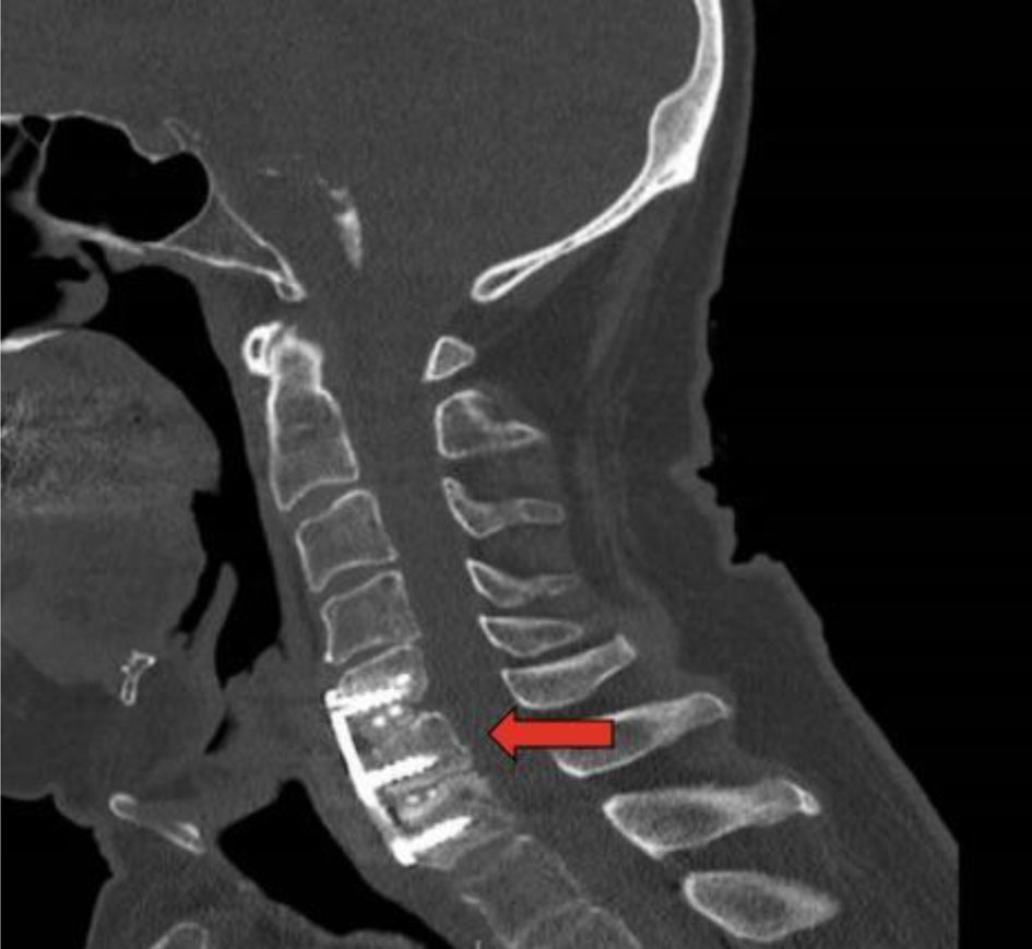
Figure 1. Computed tomography of cervical spine 6 months postoperatively. Red arrow indicates cervical plate.
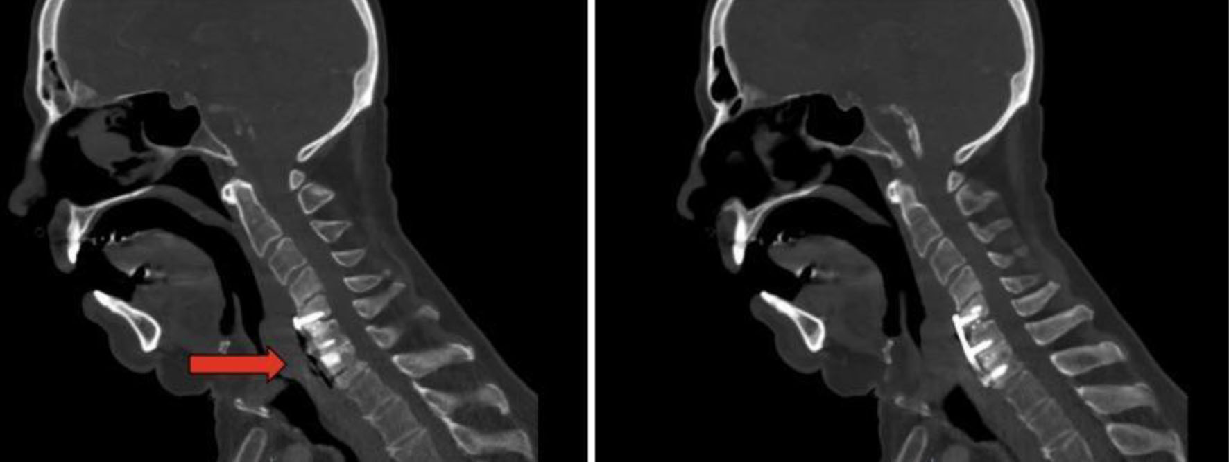
Figure 2. Computed tomography of the neck 12 months postoperatively. Anterior cervical plate in adequate position with air between the plate and esophagus.
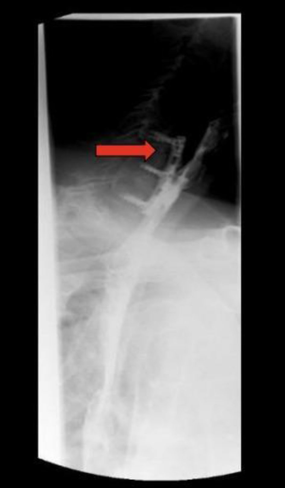
Figure 3. Esophagram at 12 months. Red arrow indicates defect. The image shows contiguity between the lumen of the esophagus and the cervical hardware based on the transit of the oral contrast, consistent with a posterior esophageal wall perforation contained by the cervical hardware without extravasation.
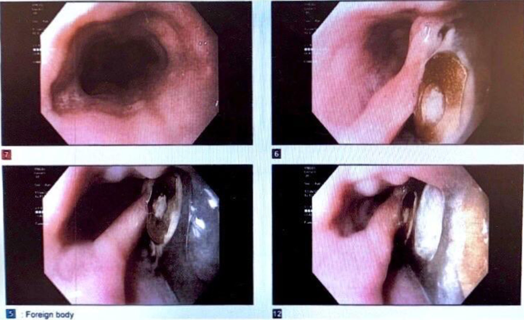
Figure 4. Esophagogastroduodenoscopy 12 months postoperatively. Image displays the cervical hardware, which had eroded through the posterior wall of the cervical esophagus concordant with the esophagram, which had demonstrated contiguity between the lumen of the esophagus and the cervical hardware based on the transit of the oral contrast, consistent with a posterior esophageal wall perforation contained by the cervical hardware without extravasation. These findings were also consistent with the patient’s chronic presentation without sepsis.
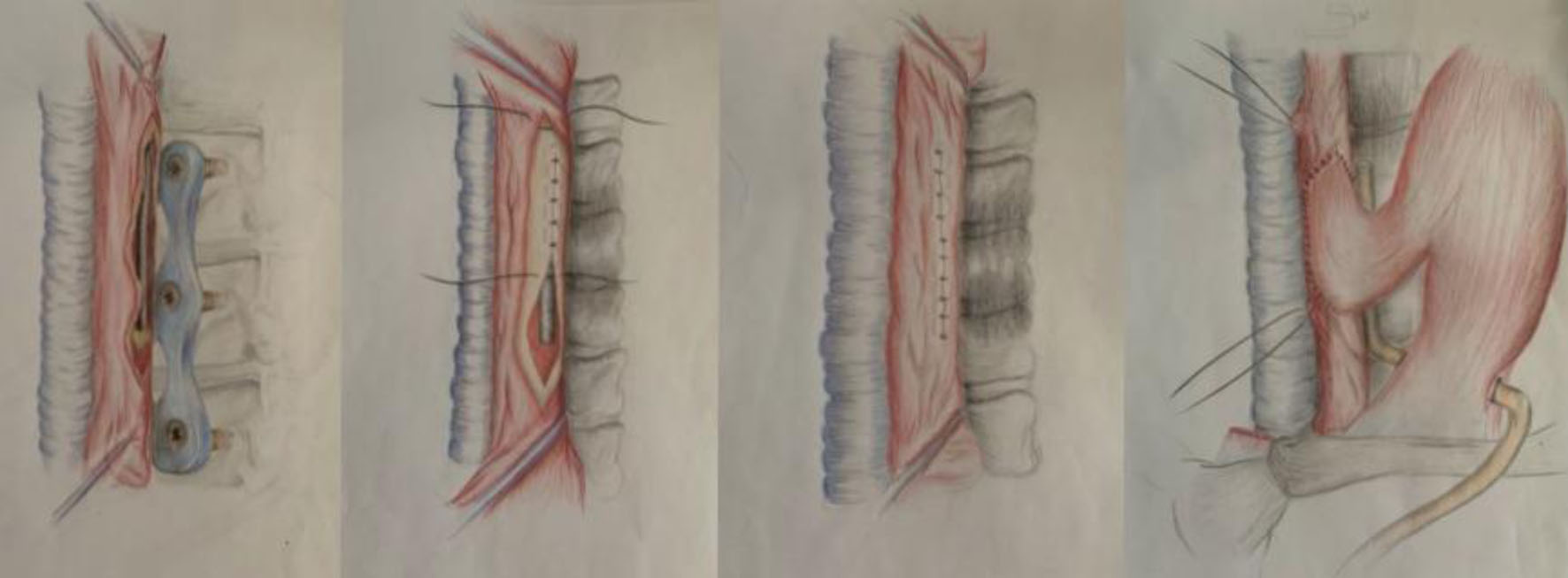
Figure 5. Representation of surgical repair guided by esophagogastroduodenoscopy in layers and muscle flap reinforcement.
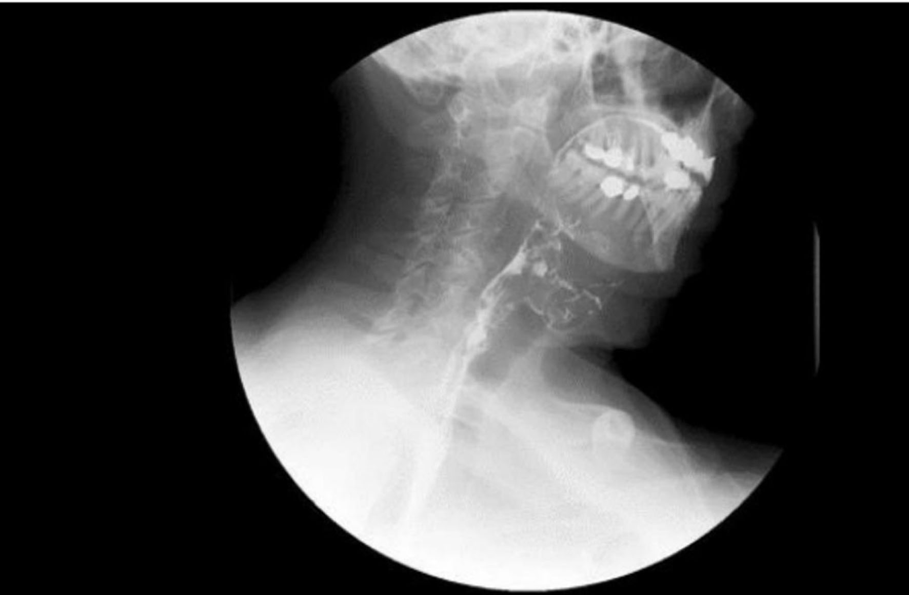
Figure 6. Postoperative barium swallow demonstrating resolution of obstruction.