World Journal of Oncology, ISSN 1920-4531 print, 1920-454X online, Open Access
Article copyright, the authors; Journal compilation copyright, World J Oncol and Elmer Press Inc
Journal website https://www.wjon.org

Case Report

Volume 14, Number 6, December 2023, pages 584-588


Borderline Phyllodes Tumor in a Child

Figures

Figure 1.
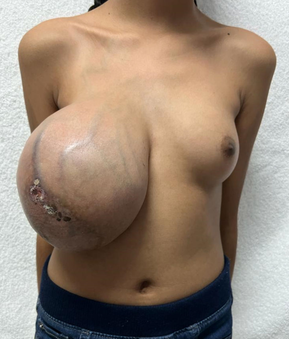
Figure 1. Large right breast mass with multiple collateral veins and ulceration of the nipple. Note the normal left breast.
Figure 2.
Figure 2. Macroscopic examination of mastectomy shows multinodular mass with pseudocystical and mucoid aspect and 20 cm of diameter.
Figure 3.
Figure 3. (a) H&E stain (× 10) shows stroma with nodular enlargement and a leaf-like pattern (arrows). (b) IHC estrogen receptor (× 20) stain shows positive immunoreactivity in 80% of malignant cells. (c) IHC progesterone receptor stain (× 20) shows positive immunoreactivity in 70% of malignant cells. (d) HER2 stain (× 20) shows no immunoreactivity. (e) Ki-67 stain (× 10) shows less than 2% of nuclear immunoreactivity on epithelial cells. IHC: immunohistochemistry; H&E: hematoxylin and eosin; HER: human epidermal growth factor receptor.