Figures
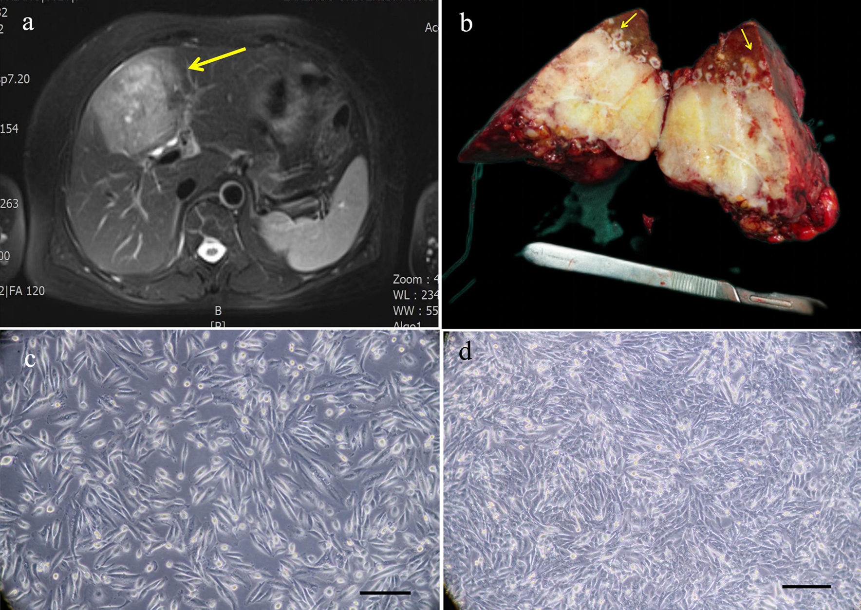
Figure 1. Relevant clinical data and morphology of the ICC-X2 cells. (a) MRI scan showing a large 9 × 7 cm lesion in segment IV of the liver (long arrow). (b) Gross view of the surgically resected specimen. Multiple satellite lesions can be observed around the primary lesion (short arrows). (c) Light microscope image of the ICC-X2 cells of passage 10. (d) Light microscope images of the ICC-X2 cells of passage 50 (scale bar = 100 µm). MRI: magnetic resonance imaging.
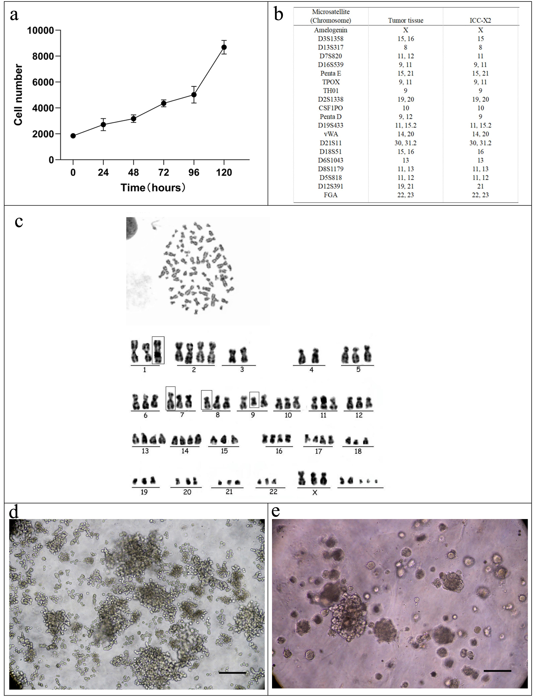
Figure 2. Cell growth curve, karyotyping analysis, DNA STR analysis, tumor sphere, and organoid culture of the ICC-X2 cells. (a) Growth curve of the ICC-X2 cells. The doubling time of the ICC-X2 cells is 48 h. (b) Representative karyotype of the ICC-X2 cells. The cells exhibit hypotetraploid karyotypes with large differences in chromosome number and morphology. (c) STR analysis indicates that the ICC-X2 cells are highly consistent with the primary tumor tissue. (d) Two-week tumor sphere culture of ICC-X2. (e) Two-week ICC-X2 organoid culture (scale bar = 100 µm). STR: short tandem repeat.
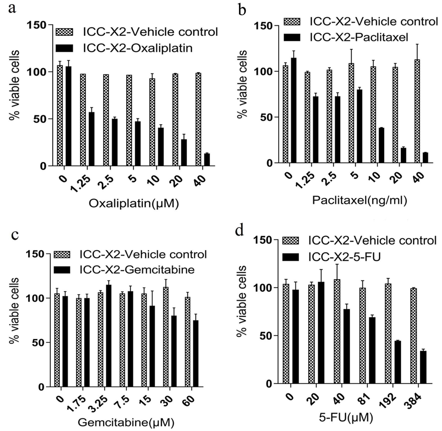
Figure 3. Drug sensitivity of the ICC-X2 cells. Dose-dependent effects of (a) paclitaxel, (b) gemcitabine, (c) oxaliplatin, and (d) 5-FU in the ICC-X2 cells. The cell line was sensitive to oxaliplatin and naturally resistant to gemcitabine, paclitaxel, and 5-FU. 5-FU: 5-fluorouracil.
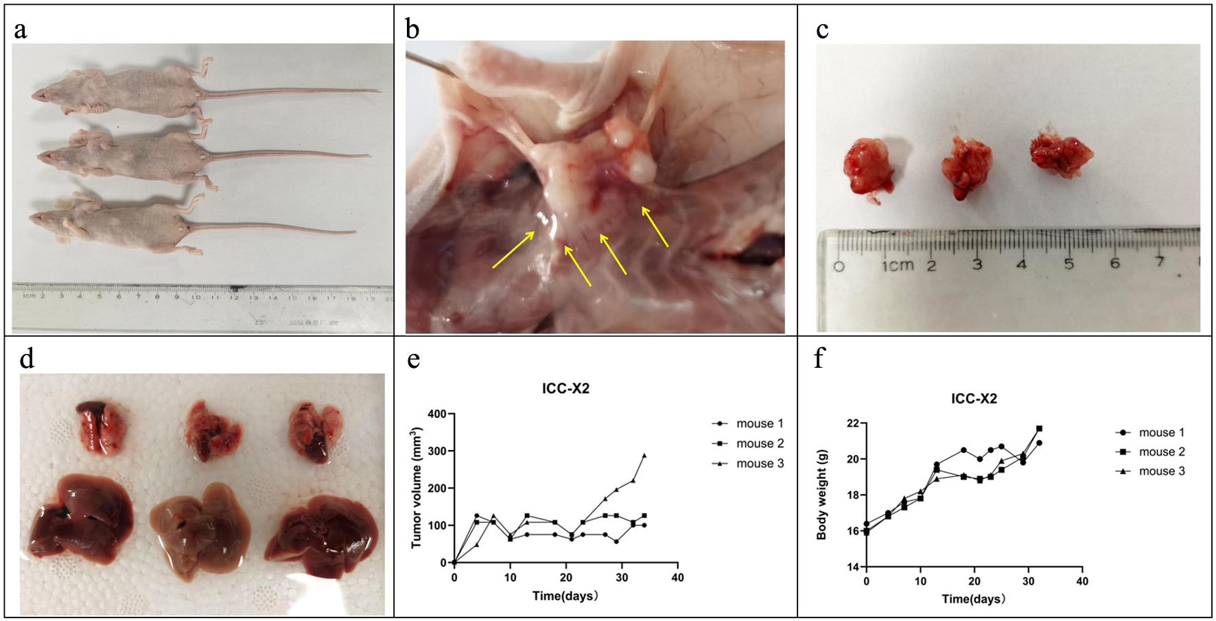
Figure 4. Tumorigenicity in BALB/C nude mice. (a) ICC-X2 cells can rapidly form xenografts after inoculation into BALB/C nude mice. (b) ICC-X2 xenograft tumors demonstrate stronger peritumoral infiltration (arrow). (c) Xenograft tumors develop rapidly, and the diameter of the tumor is close to 1 cm at 4 weeks. (d) No metastatic lesions are observed in other organs of the transplanted mice within 4 weeks. (e) Tumor volume curve when the ICC-X2 cells were inoculated into BALB/C nude mice. (f) Weight curve when the ICC-X2 cells were inoculated into BALB/C nude mice.
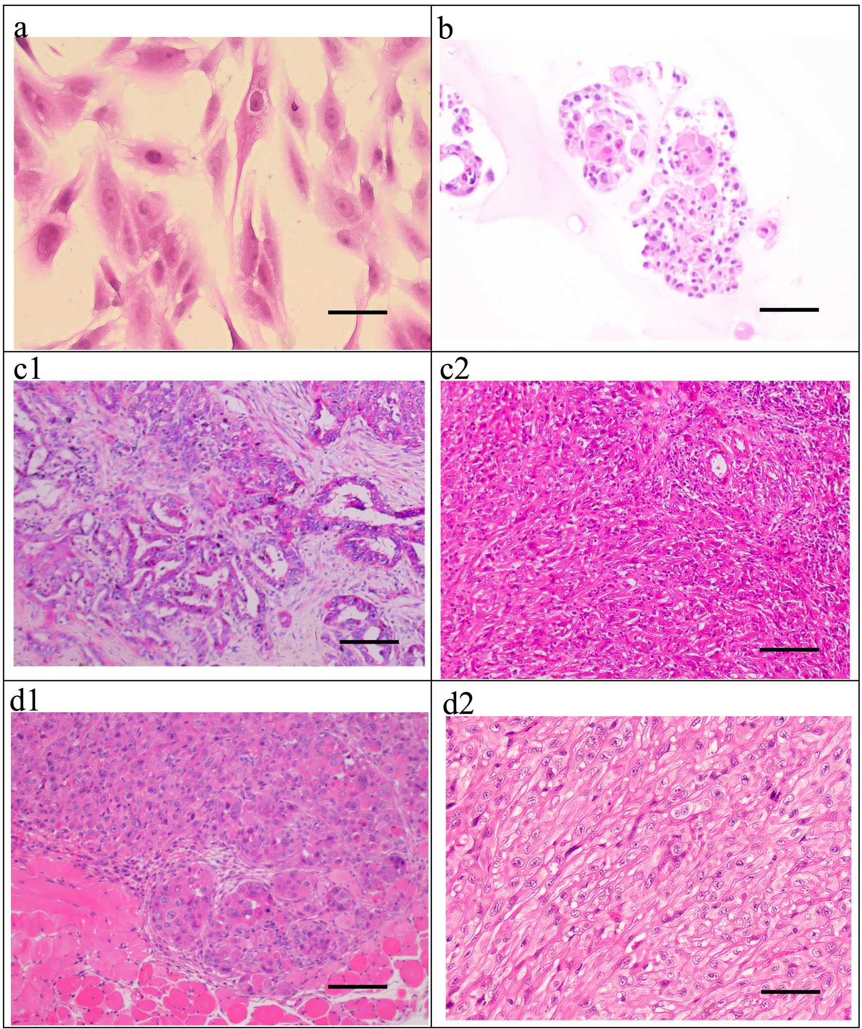
Figure 5. H&E staining of the ICC-X2 cells, organoids, primary tumor, and xenograft tumor. (a) H&E staining of the ICC-X2 cells. (b) H&E staining of the ICC-X2 organoids. H&E staining of the primary tumor shows moderately to poorly differentiated ICC, and the cancer cells are mainly polygonal and round (c1), some of which are undifferentiated cells, as indicated by their spindle-shaped appearance (c2). H&E staining of the xenograft tumors demonstrates that some cells form gland-like structures (d1), while some cells are spindle-shaped (d2) similar to fibroblasts (scale bar = 50 µm). H&E: hematoxylin and eosin.
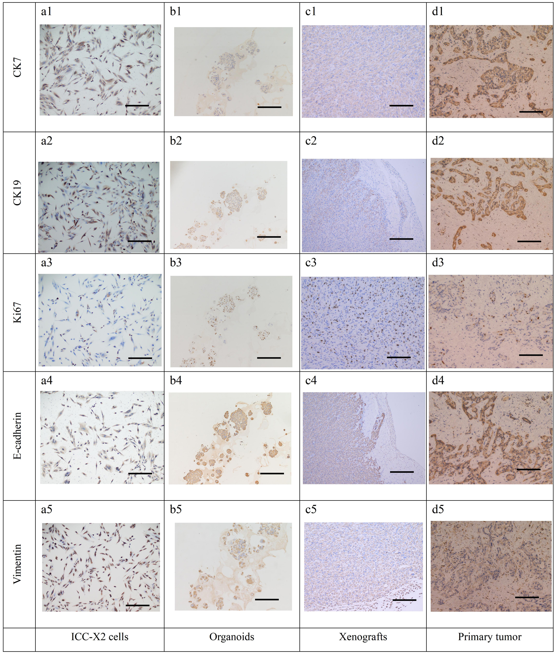
Figure 6. IHC staining of the cells, organoids, xenograft, and the primary tissue of ICC-X2. CK7 positive staining in cells (a1), organoids (b1), xenograft (c1), and the primary tissue (d1) of ICC-X2. CK19 positive staining in cells (a2), organoids (b2), xenograft (c2), and the primary tissue (d2) of ICC-X2. Ki-67 positive staining in cells (a3), organoids (b3), xenograft (c3), and the primary tissue (d3) of ICC-X2. E-cadherin positive staining in cells (a4), organoids (b4), xenograft (c4), and the primary tissue (d4) of ICC-X2. Vimentin positive staining in cells (a5), organoids (b5), xenograft (c5), and the primary tissue (d5) of ICC-X2 (scale bar = 100 µm). IHC: immunohistochemical.
Table
Table 1. Clinical Data of the Patient Included in the Study
| Cell line | Patient age/ethnicity | Gender | Current status (days) | Histopathology/differentiation | Tumor size (cm) | Prior therapy | Culture date | Microvascular invasion | Nerve invasion | Lymph node and distant metastasis | AJCC cancer staging | Serum |
|---|
| AFP (0 - 5.8 IU/mL) | CEA (0 - 5.2 ng/mL) | CA19-9 (0 - 35 U/mL) |
|---|
| AJCC: American Joint Committee on Cancer; AFP: alpha-fetoprotein; CEA: carcinoembryonic antigen; CA19-9: carbohydrate antigen 19-9. |
| ICC-X2 | 62/Asian | Female | NA | Moderately-poorly, some undifferentiated | 9 × 7 | None | July 23, 2021 | No | Yes | Nos. 12 lymph nodes (2/2) and Nos. 13 lymph nodes (5/5); abdominal wall metastasis and omental metastasis | T2N1M0 | 7.8 | 1.7 | 24.9 |