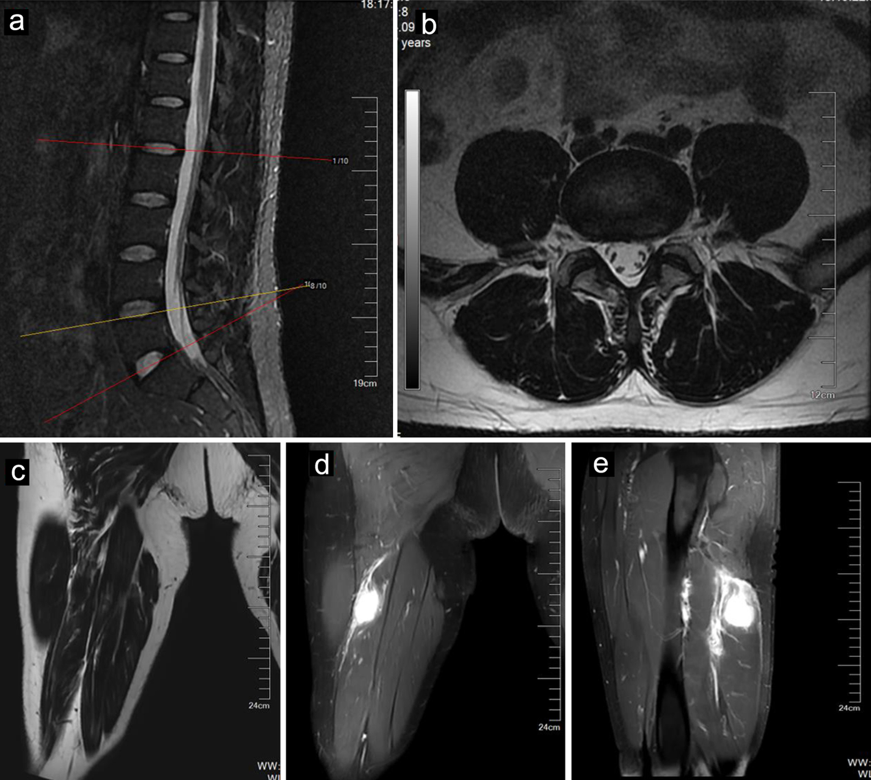
Figure 1. MR images of a 40-year-old male with IMT. Sagittal (a) and axial (b) MR images of the lumbar spine showing a mildly herniated disc at the L4-L5 level. The tumor (arrows) showed mild hyperintensity on T1-weighted imaging (c) and hyperintensity on fat-suppressed T2-weighted imaging (d). The tumor (arrows) showed enhancement after contrast agent injection (e). IMT: inflammatory myofibroblastic tumor; MR: magnetic resonance.
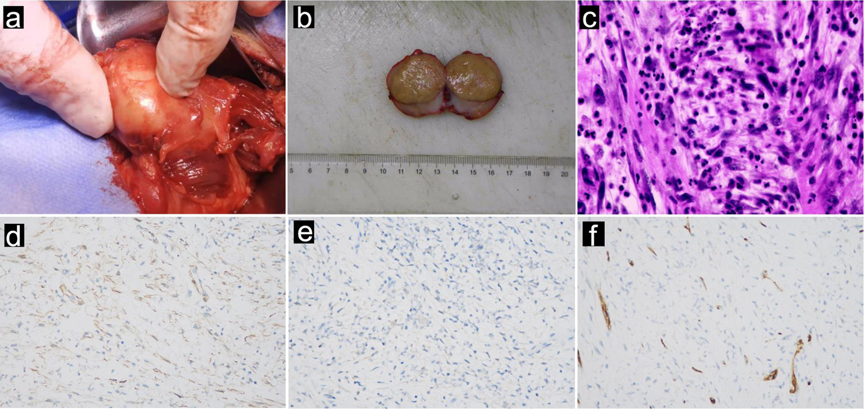
Figure 2. Intraoperative and pathological images of a 40-year-old male with IMT derived from the sciatic nerve. (a) Intraoperative photograph, (b) macroscopic photograph, and (c) microscopic examination revealing IMTs. The tumor cells consisted of fusiform myofibroblasts, infiltrated plasma cells, lymphocytes, and eosinophils. The tumor cells did not express SMA (d), original magnification × 400; S100 (e), original magnification × 400, or CD34 (f), original magnification × 400, confirming the diagnosis of IMT. IMT: inflammatory myofibroblastic tumor.