World Journal of Oncology, ISSN 1920-4531 print, 1920-454X online, Open Access
Article copyright, the authors; Journal compilation copyright, World J Oncol and Elmer Press Inc
Journal website https://www.wjon.org

Review

Volume 15, Number 3, June 2024, pages 372-381


Comprehensive Insights Into Renal Perivascular Epithelioid Cell Neoplasms: From Molecular Mechanisms to Clinical Practice

Figures

Figure 1.
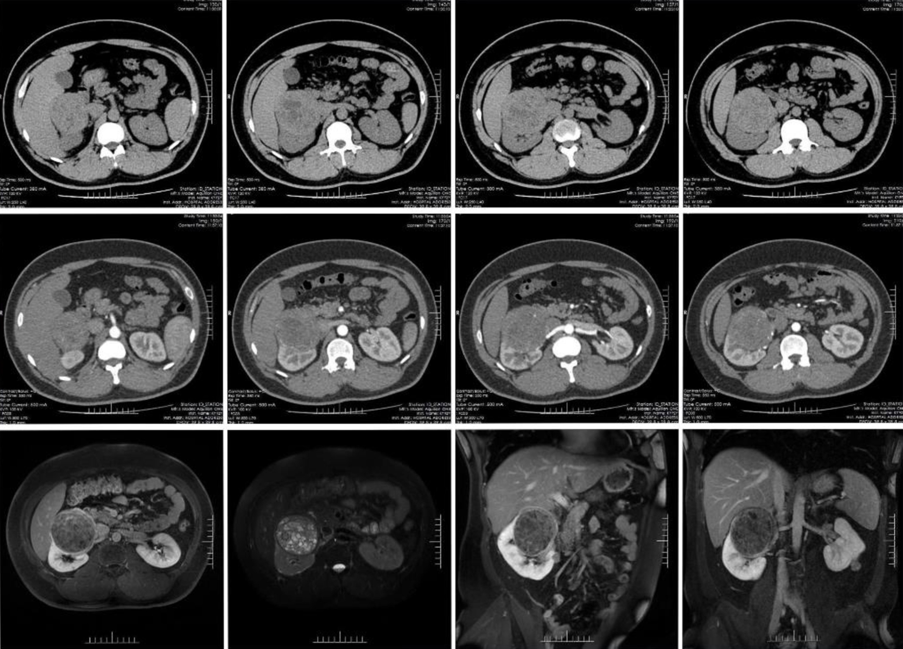
Figure 1. The computed tomography (CT) and magnetic resonance imaging (MRI) of the patient.
Figure 2.
Figure 2. The radical nephrectomy of the right kidney. (a) Adhesion of the right kidney to the descending portion of the duodenum. (b) The kidney and the duodenum after adhesion were released. (c) Adhesion of the right kidney to the inferior vena cava. (d) The kidney and the inferior vena cava after adhesion were released. (e) The lower lobe of the liver after adhesion was released.
Figure 3.
Figure 3. The histopathology (a-e) and immunohistochemistry (f-h) of the right renal mass. (f) HMB45 positive, (g) melan-A positive, (h) TFE3 negative.
Figure 4.
Figure 4. The case history.