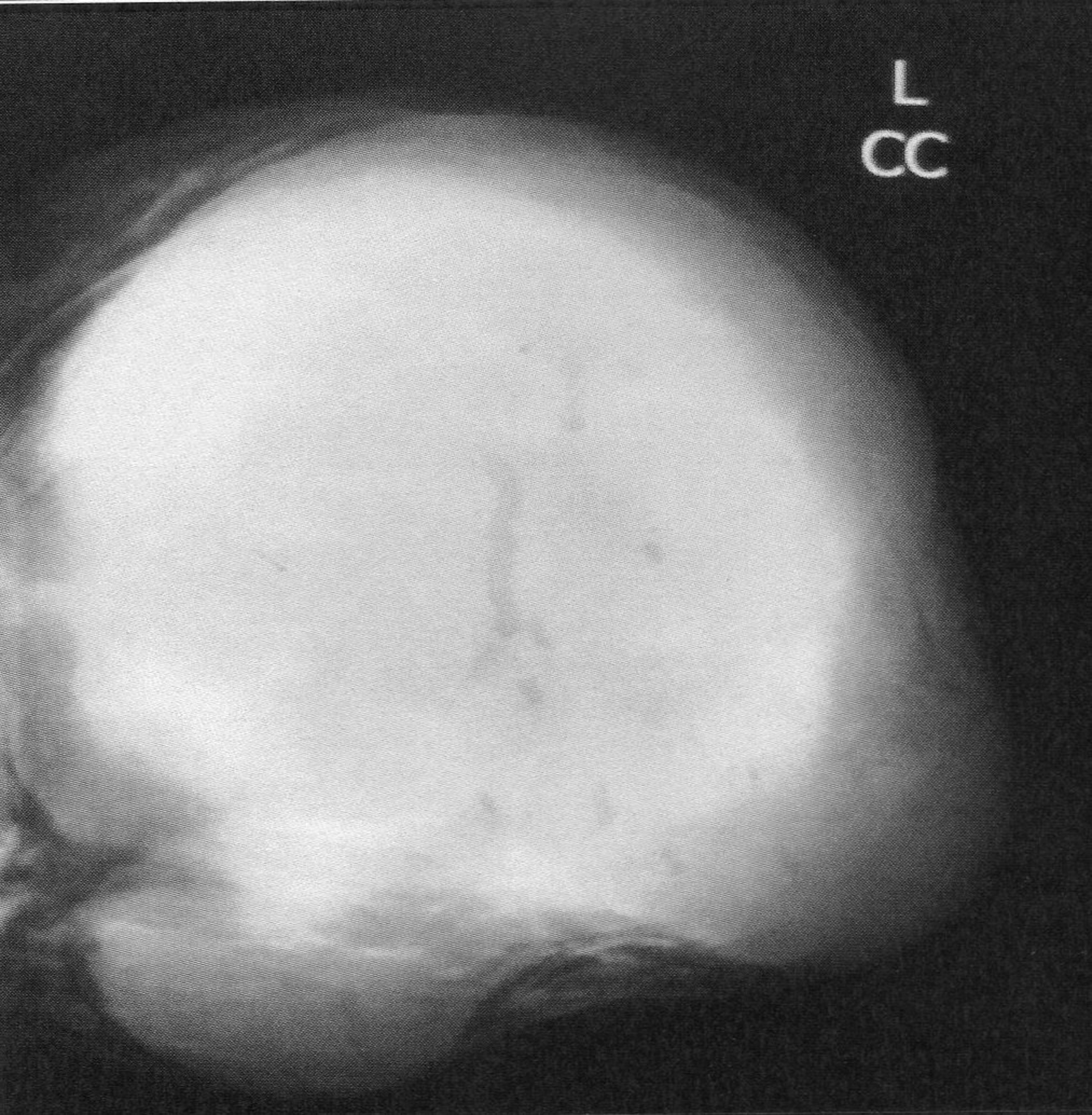
Figure 1. Mammography revealing a lobulated mass with smooth margins.
| World Journal of Oncology, ISSN 1920-4531 print, 1920-454X online, Open Access |
| Article copyright, the authors; Journal compilation copyright, World J Oncol and Elmer Press Inc |
| Journal website http://www.wjon.org |
Original Article
Volume 1, Number 3, June 2010, pages 129-134
Phyllodes Tumors of the Breast: A Review of 26 Cases
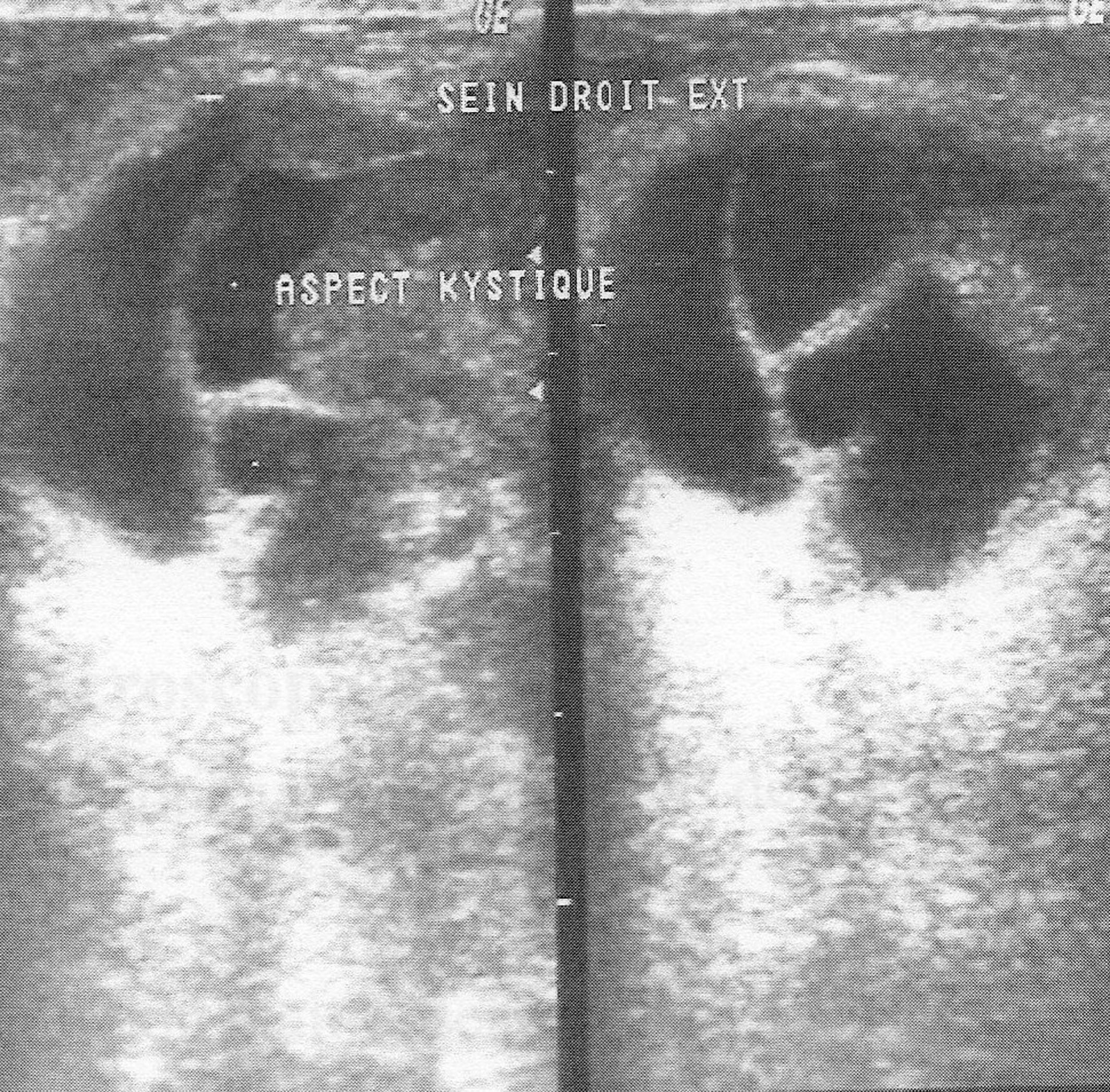
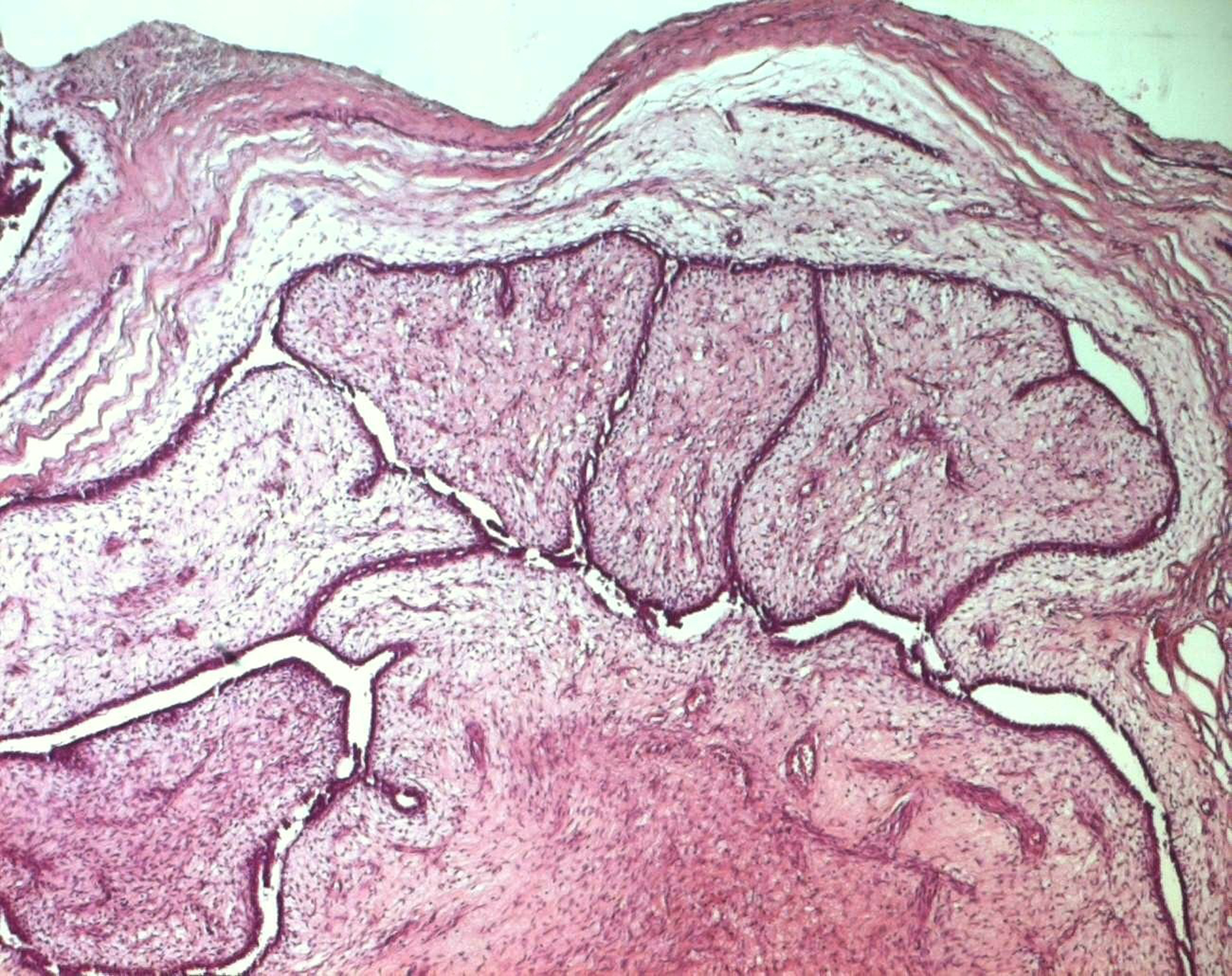
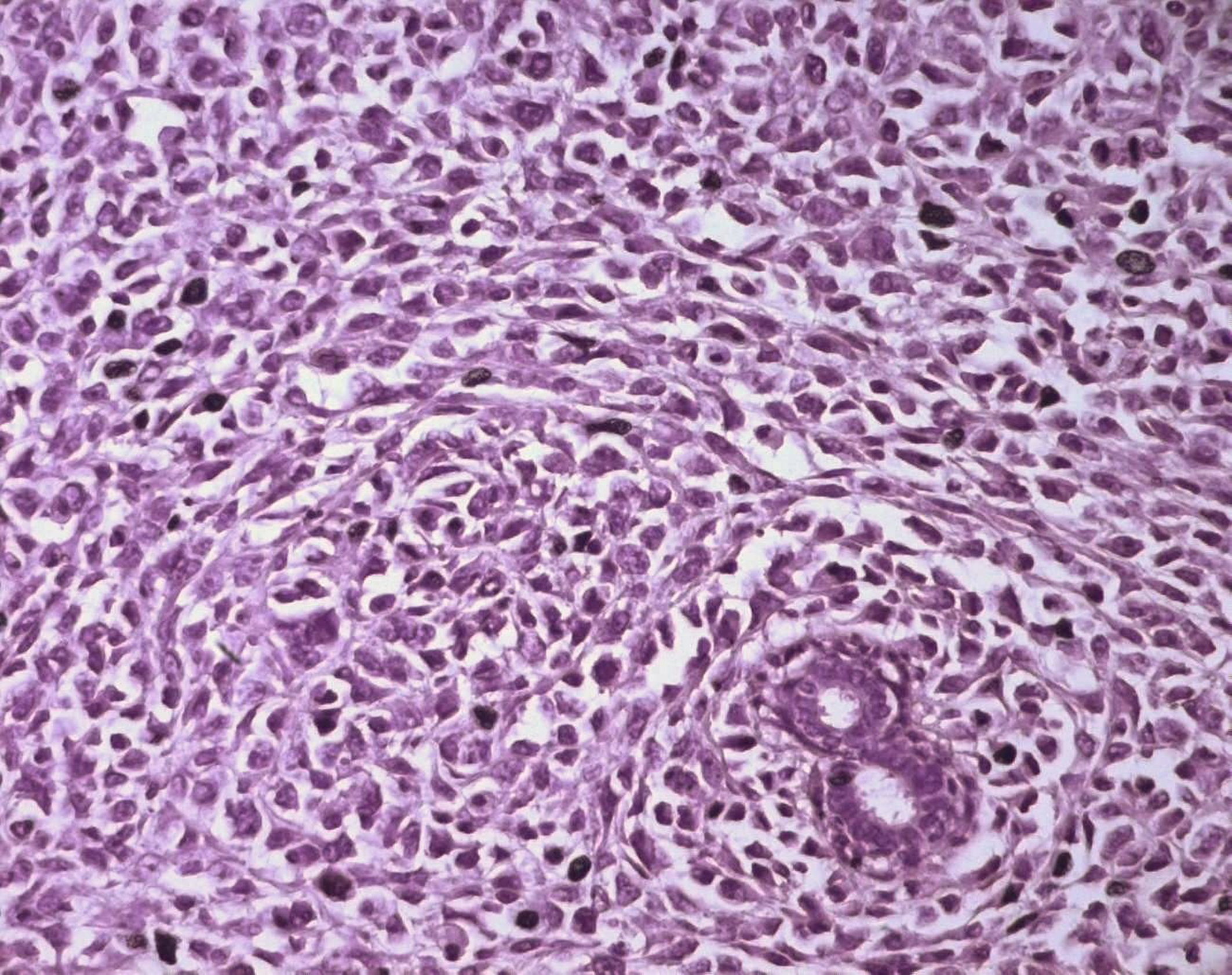
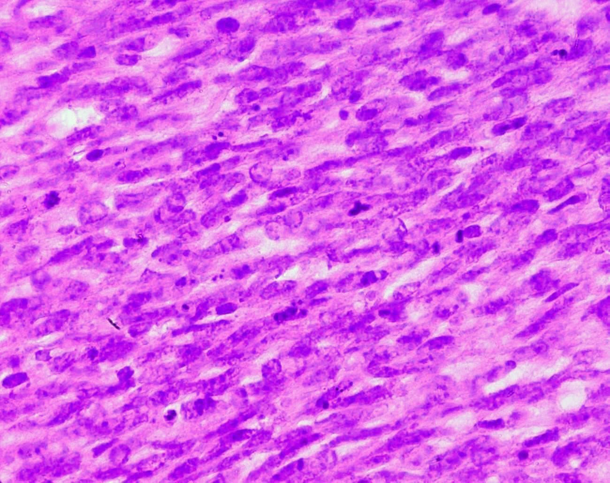
Figures
Table
| Benign | Borderline | Malignant | |
|---|---|---|---|
| *One patient developed recurrent disease at 10 months and lung metastases at 21 months. | |||
| Number of patients | 13 | 7 | 6 |
| Mean age (range) | 35.8 (19 - 55) | 44.7 (34 - 52) | 45.2 (29 - 66) |
| Size | |||
| < 5 cm | 6 | 2 | 3 |
| ≥ 5 cm | 7 | 5 | 3 |
| Surgical approach | |||
| breast-conserving surgery | 12 | 6 | 2 |
| mastectomy | 1 | 1 | 4 |
| Surgical margin | |||
| negative | 6 | 4 | 4 |
| close | 5 | 3 | 2 |
| indeterminate | 2 | 0 | 0 |
| Outcome | |||
| Recurrence | 1 | 3* | 2 |
| Metastases | 0 | 1* | 2 |
| Favorable | 10 | 4 | 0 |
| Lost to follow-up | 2 | 0 | 2 |