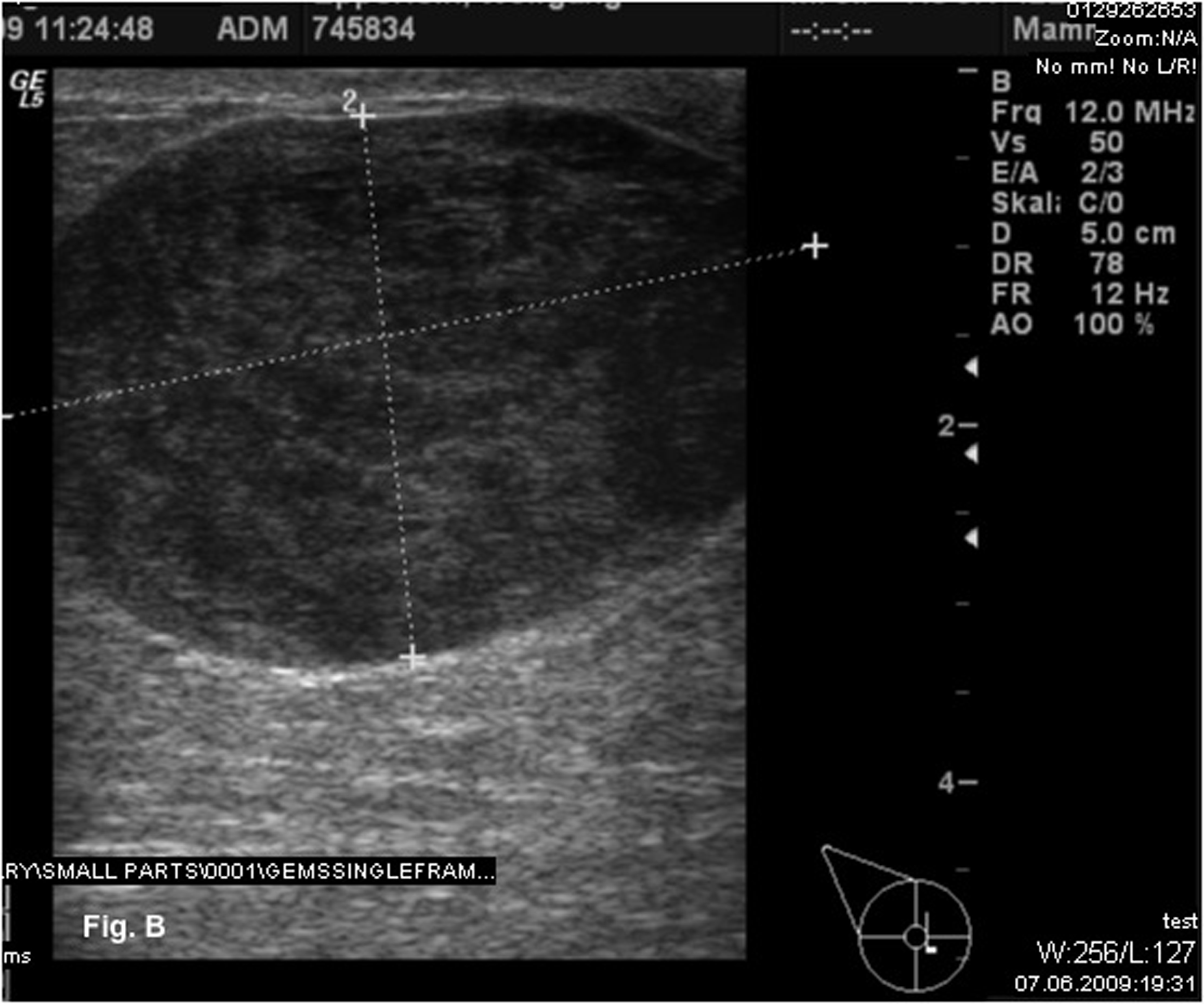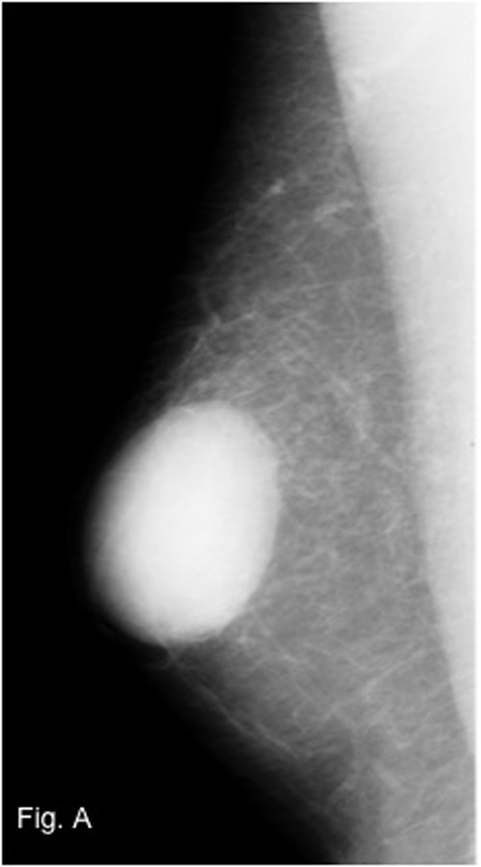
Figure 1. Mammography of the right breast with a dense mass in a central localization.
| World Journal of Oncology, ISSN 1920-4531 print, 1920-454X online, Open Access |
| Article copyright, the authors; Journal compilation copyright, World J Oncol and Elmer Press Inc |
| Journal website http://www.wjon.org |
Case Report
Volume 1, Number 5, October 2010, pages 210-212
Primary Leiomyosarcoma of the Male Breast
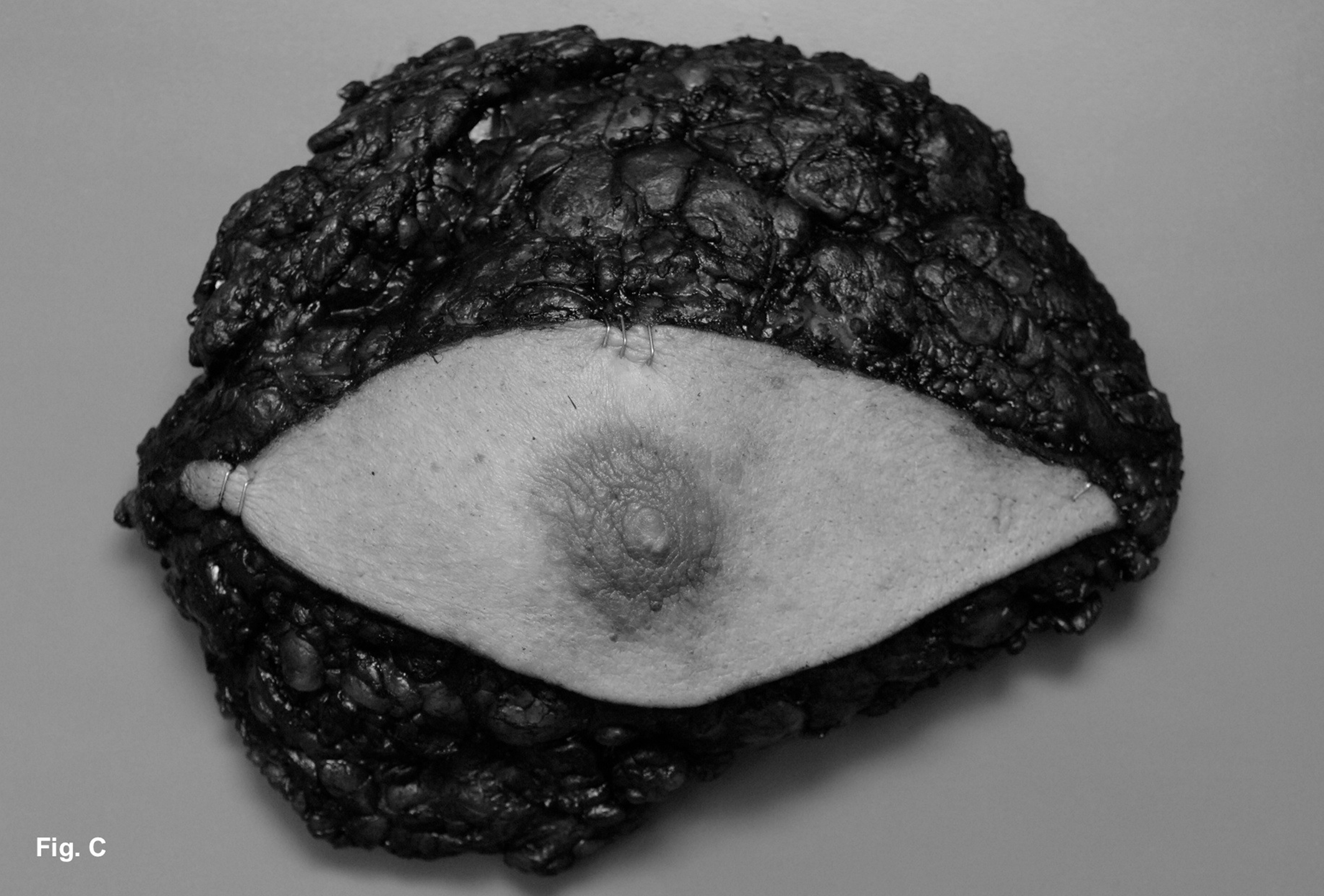
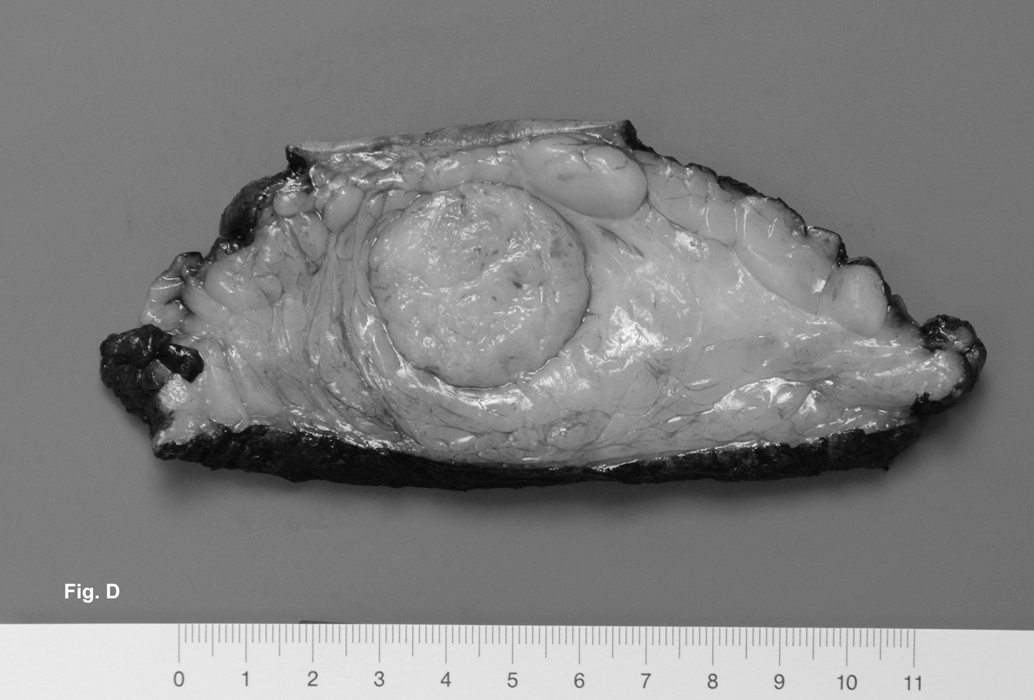
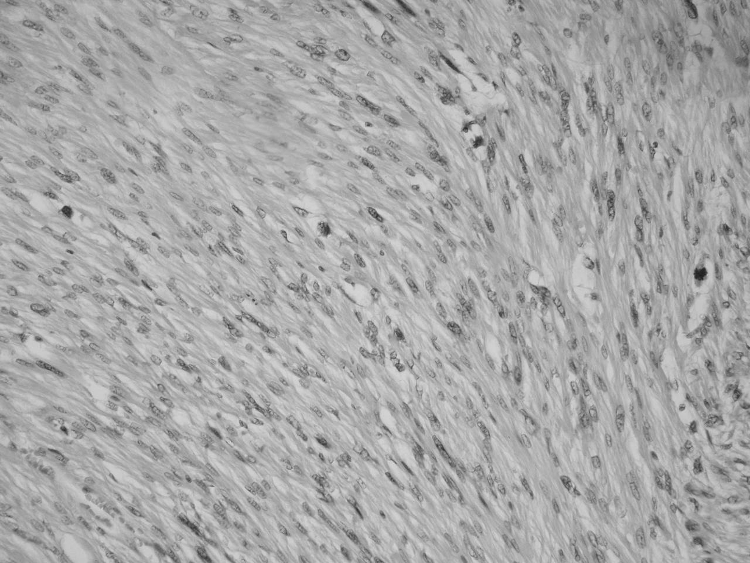
Figures