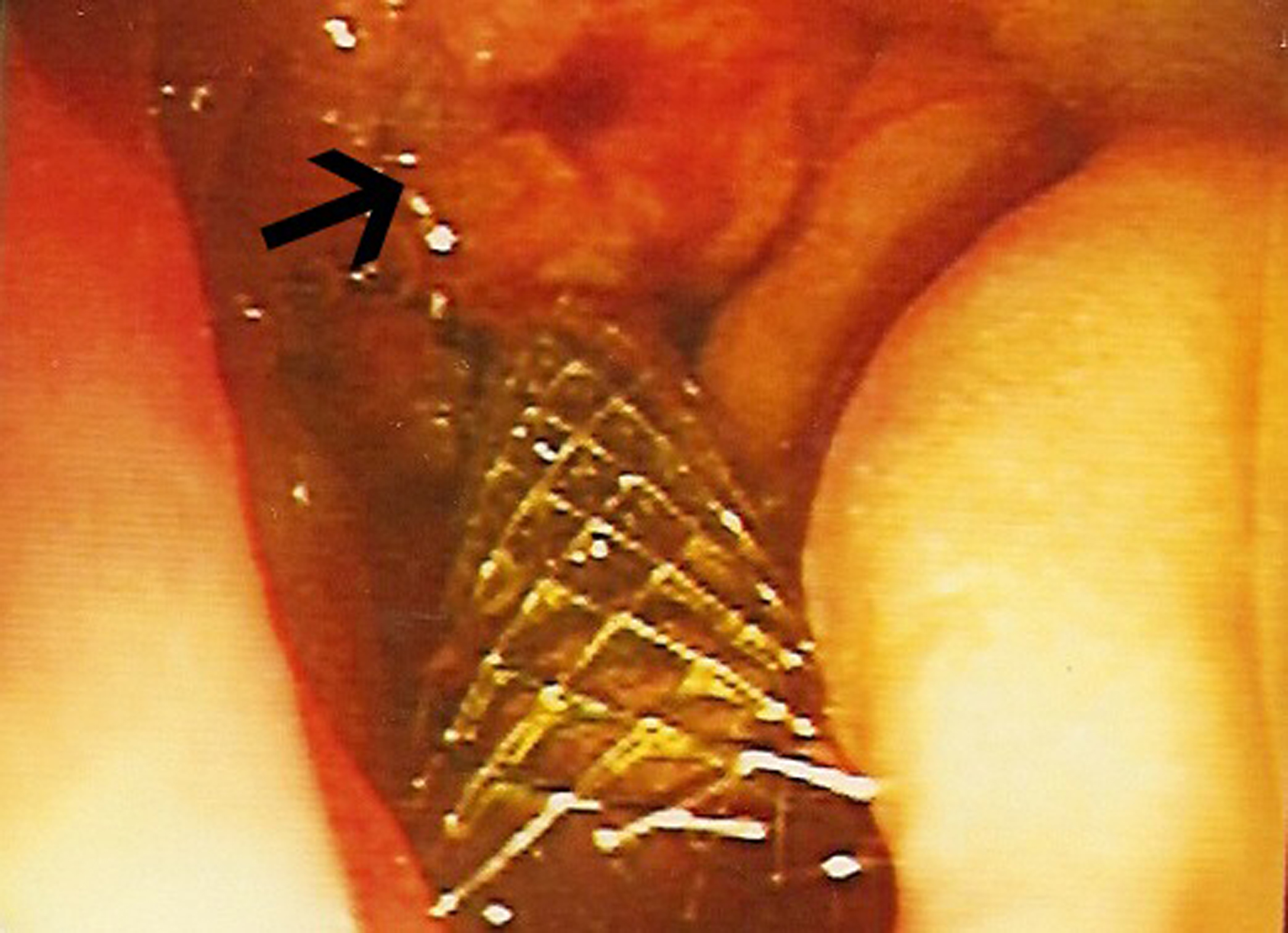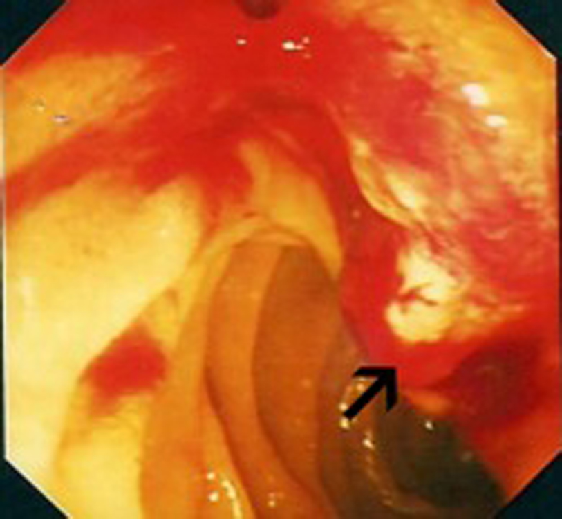
Figure 1. Endoscopic view: black arrow pointing to bleeding mass at the ampulla of Vater.
| World Journal of Oncology, ISSN 1920-4531 print, 1920-454X online, Open Access |
| Article copyright, the authors; Journal compilation copyright, World J Oncol and Elmer Press Inc |
| Journal website http://www.wjon.org |
Case Report
Volume 1, Number 5, October 2010, pages 218-220
A Case of Metastatic Renal Cell Cancer Presenting as Jaundice
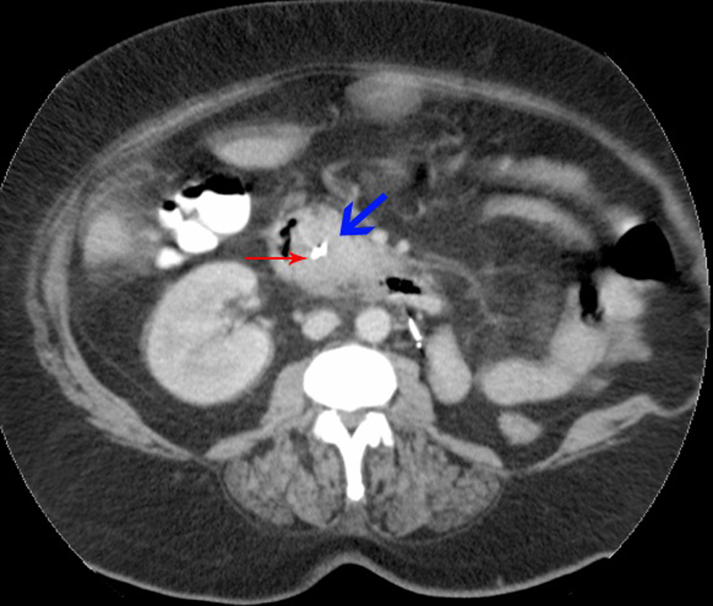
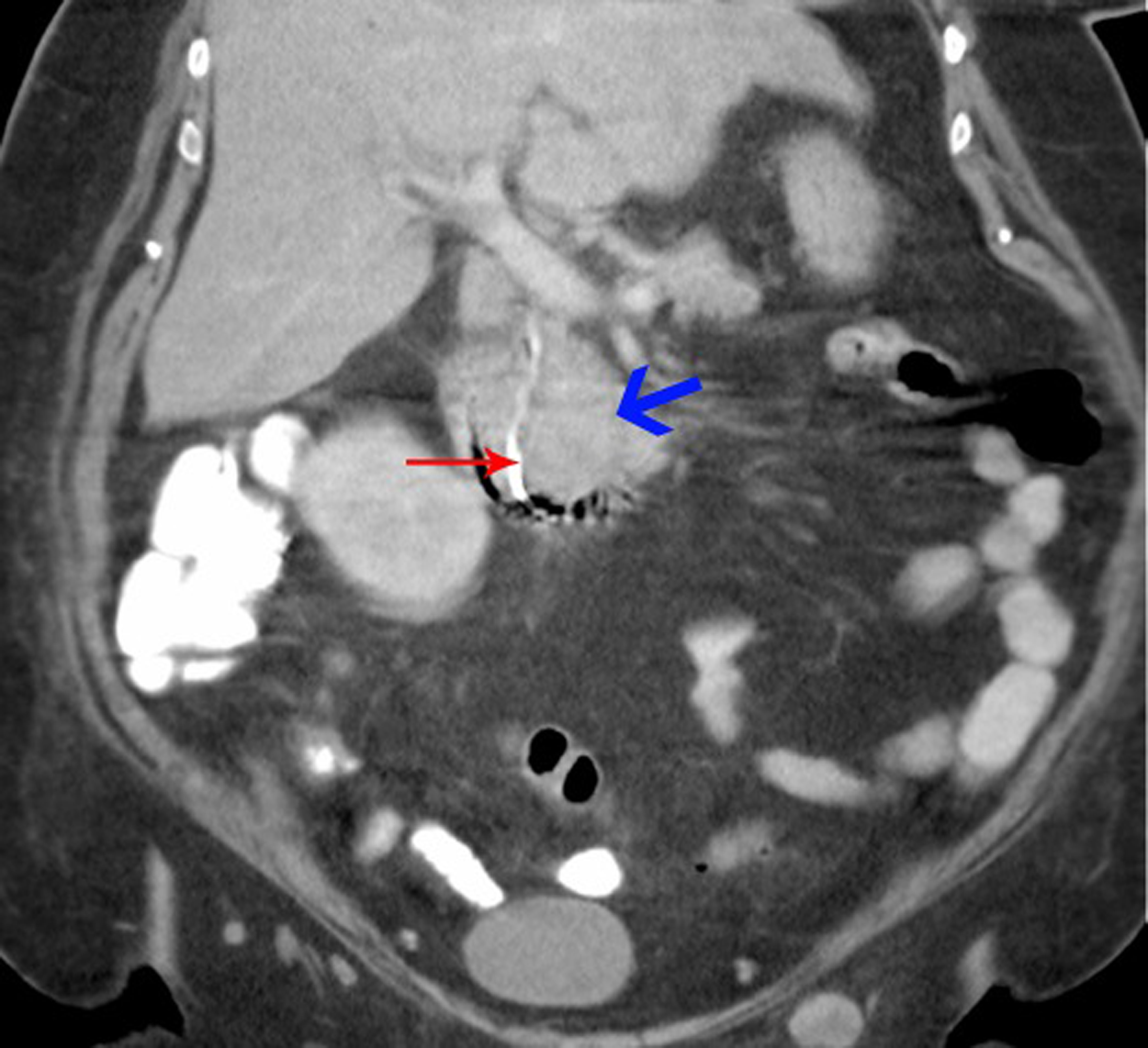
Figures