World Journal of Oncology, ISSN 1920-4531 print, 1920-454X online, Open Access
Article copyright, the authors; Journal compilation copyright, World J Oncol and Elmer Press Inc
Journal website http://www.wjon.org

Case Report

Volume 2, Number 4, August 2011, pages 195-198


Paraneoplastic Necrotizing Myelopathy in a Patient With Newly Diagnosed Diffuse Large B Cell Lymphoma

Figures

Figure 1.
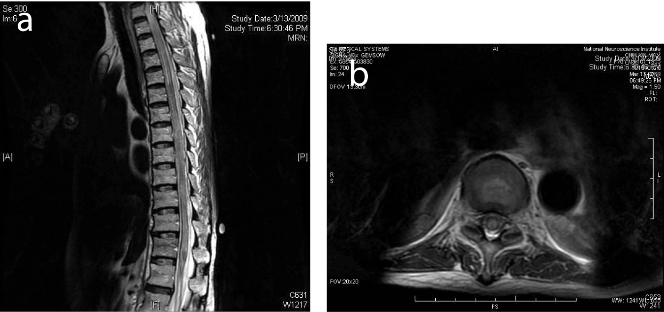
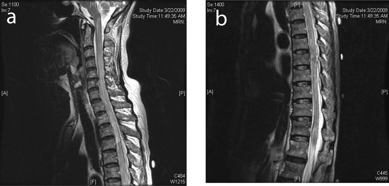
Figure 1. (a) Selected sagittal MR image of the thoracic spine on initial presentation showed normal cord signal. (b) Cervical cord on presentation showed normal MR signal.
Figure 2.
Figure 2. (a) MRI of the spine 1 week after presentation showed diffuse high T2 signal from the spinal cord from T6 to the conus compatible with cord edema. (b) Selected corresponding axial MR image showed edema across the whole cross-section of the spinal cord.
Figure 3.
Figure 3. (a) MRI of the spine showing cord edema progressing cranially to involve the cervical spine. (b) MRI demonstrated edema involving the conus medullaris.
Figure 4.
Figure 4. Selected sagittal MR image of the cervical and upper thoracic spine after treatment with methyprednisolone and resumption of chemotherapy, showed near resolution of the abnormal cord signal.