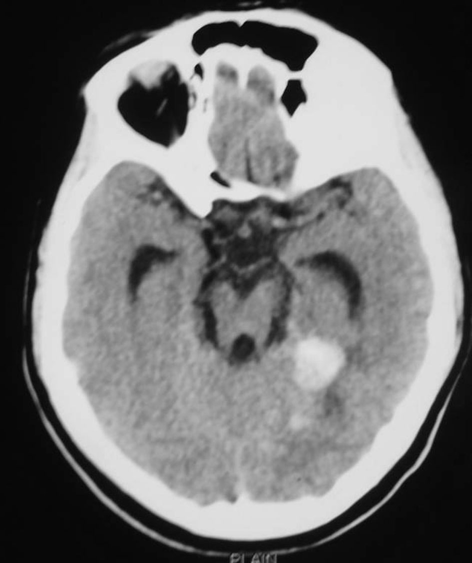
Figure 1. CT scan of the brain showing diffuse subarachnoid hemorrhage and foci of bleed in the right medial posterior temporal region.
| World Journal of Oncology, ISSN 1920-4531 print, 1920-454X online, Open Access |
| Article copyright, the authors; Journal compilation copyright, World J Oncol and Elmer Press Inc |
| Journal website http://www.wjon.org |
Case Report
Volume 2, Number 2, April 2011, pages 79-82
Malignant Course of a Metastatic Melanoma During Pregnancy: A Case Report
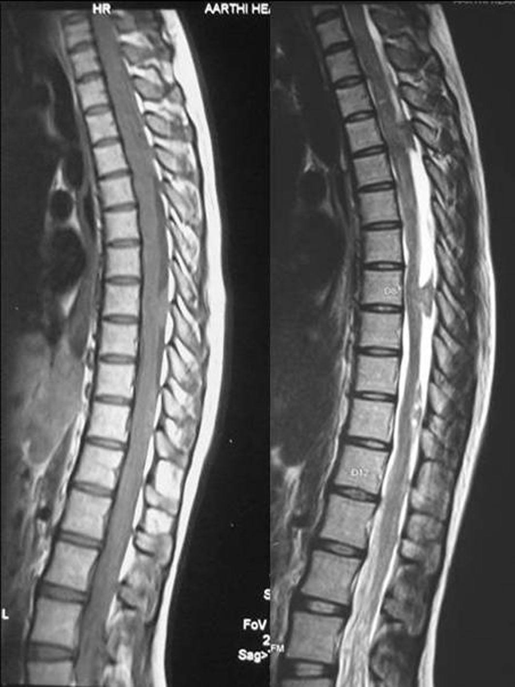
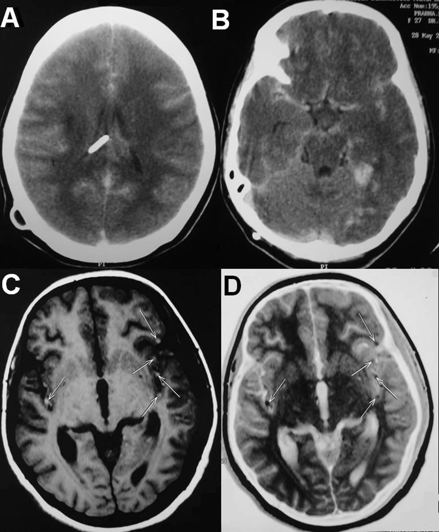
Figures