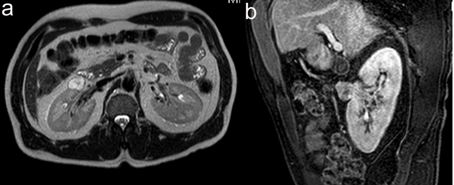
Figure 1. (a) 2.1 cm x 2.3 cm x 2.4 cm, T2 hyperintense, enhancing soft tissue renal mass anterior to interpole of right kidney. (b) Sagittal view of T1 weighted thrive sequence.
| World Journal of Oncology, ISSN 1920-4531 print, 1920-454X online, Open Access |
| Article copyright, the authors; Journal compilation copyright, World J Oncol and Elmer Press Inc |
| Journal website http://www.wjon.org |
Case Report
Volume 2, Number 2, April 2011, pages 85-88
Renal Inflammatory Myofibroblastic Tumor: A Case Report and Comprehensive Review of Literature
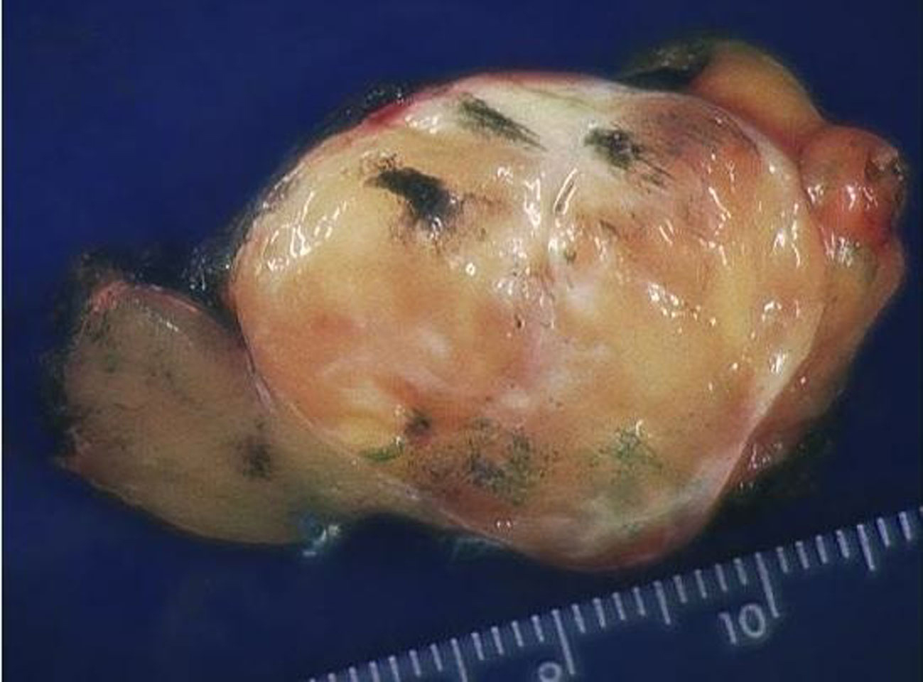
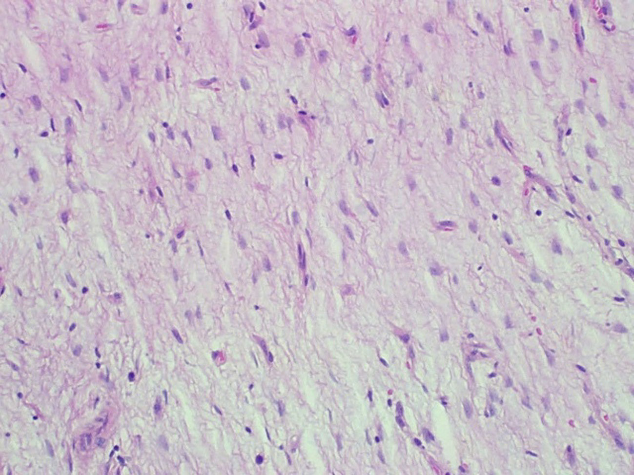
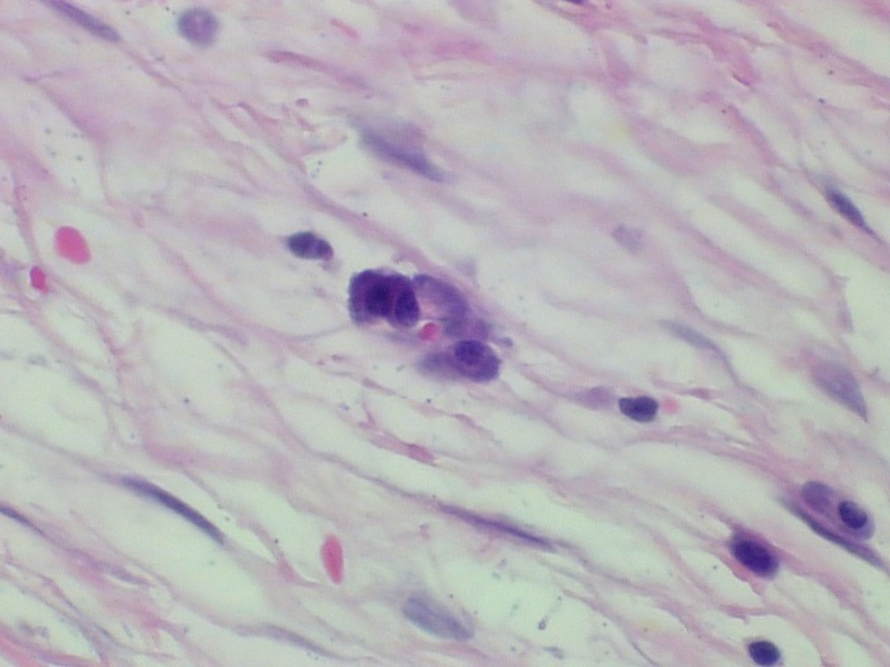
Figures