Figures
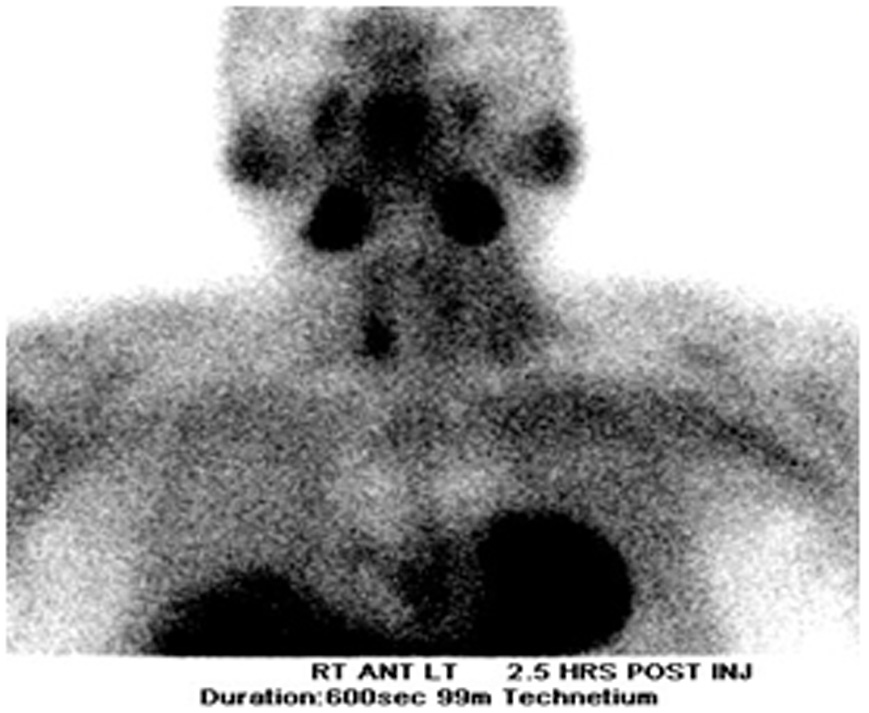
Figure 1. Parathyroid scan
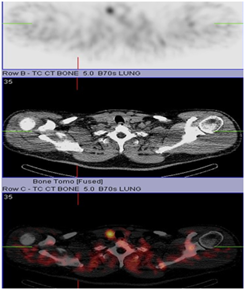
Figure 2. CT/SPECT
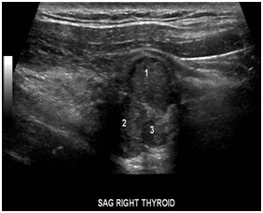
Figure 3. Ultrasound showing parathyroid mass in relation to the two thyroid nodules.
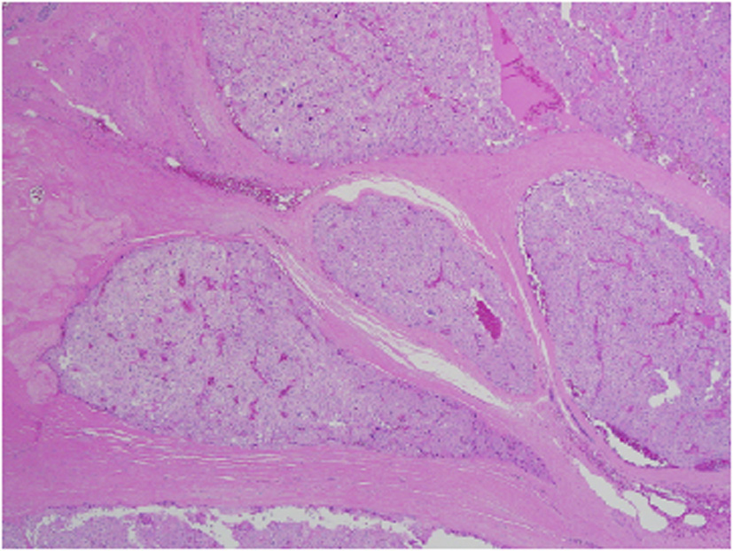
Figure 4. Low power view of parathyroid carcinoma highlighting thick surrounding and intersecting fibrous bands.
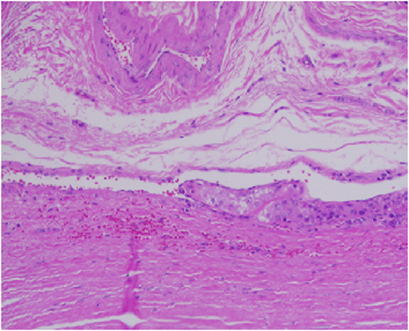
Figure 5. This focus is highly suspicious for invasion of an adjacent vessel by parathyroid carcinoma.
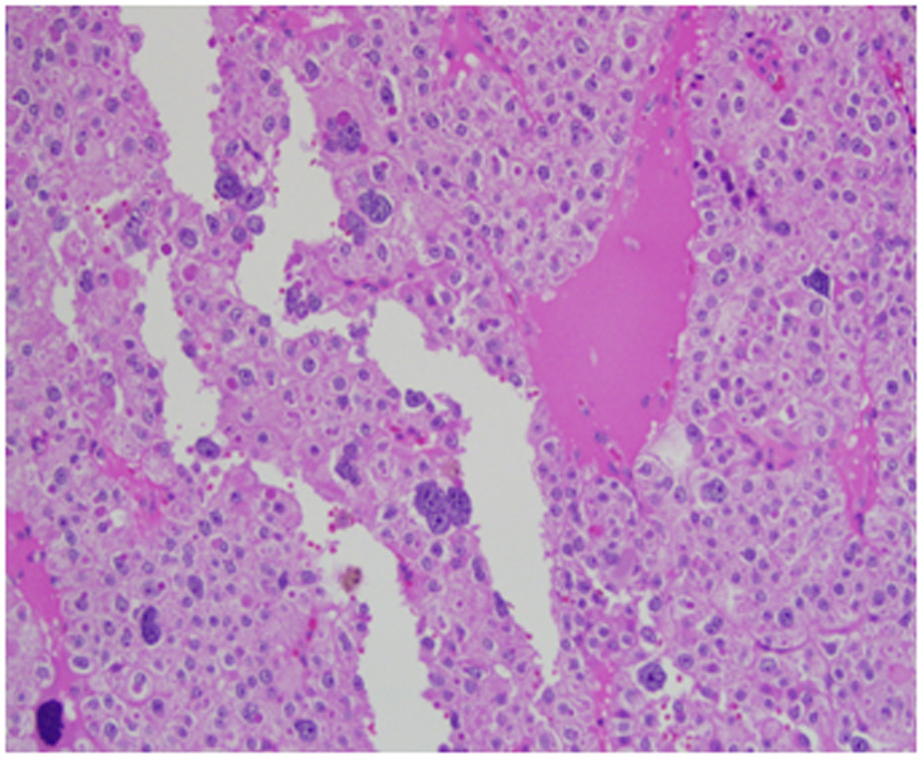
Figure 6. High power view of parathyroid carcinoma highlighting marked nuclear pleomorphism with atypical cells, prominent nucleoli, multinucleation, and abundant droplet filled cytoplasm. The chromatin varies from coarse and clumped in overtly malignant cells with marked pleomorphism to finely stippled (“salt and pepper”) in smaller more typical parathyroid tissue.
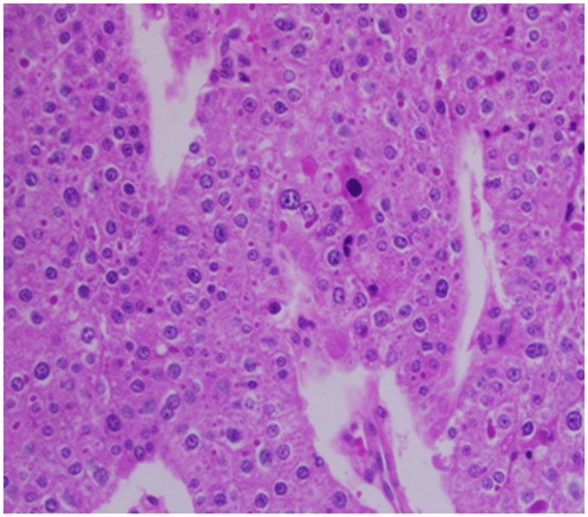
Figure 7. Prominent and irregular eosinophilic nucleoli are seen in this 40 x field with focal perinucleolar halos. The N : C ratios are moderately increased above what is typically seen in parathyroid tissue.
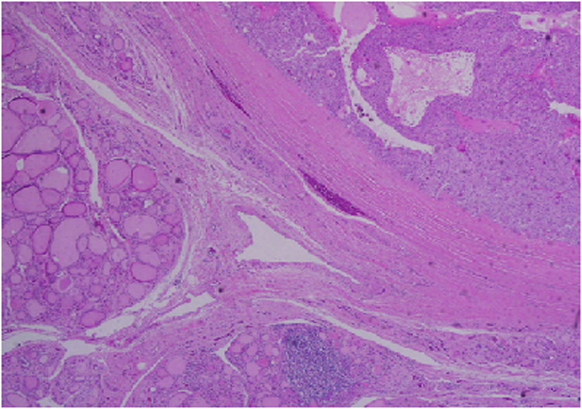
Figure 8. Low power view showing parathyroid carcinoma (upper right) and adjacent thyroid tissue (lower left).
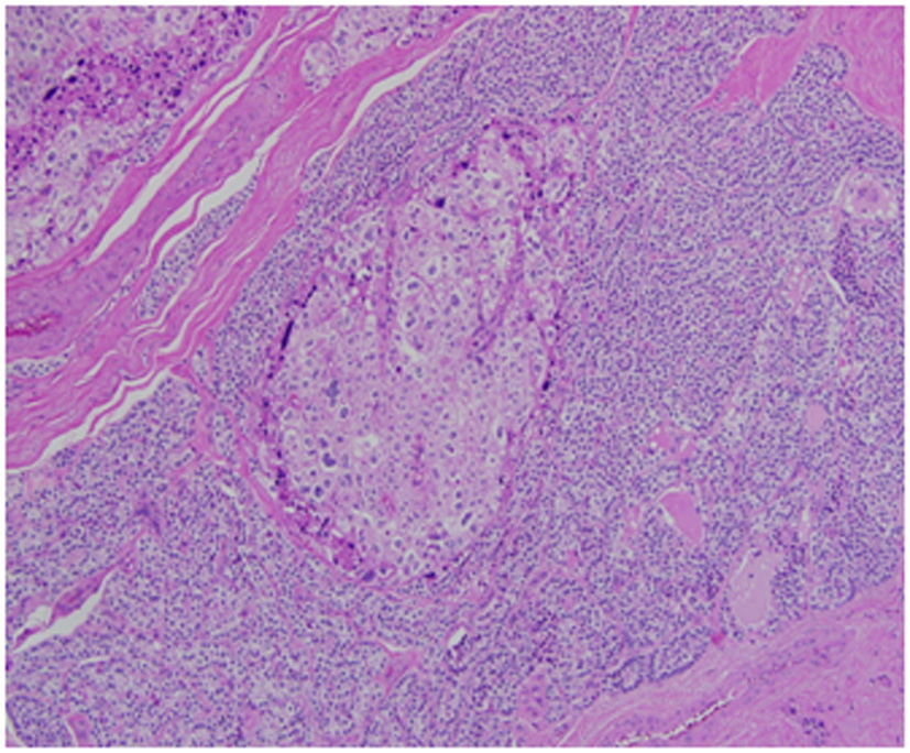
Figure 9. Medium power view of parathyroid carcinoma showing marked nuclear pleomorphic in the background of more typical parathyroid tissue.