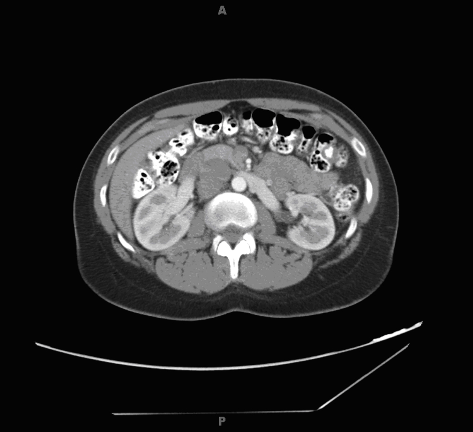
Figure 1. Abdominal computed tomography scan showing right paracaval mass.
| World Journal of Oncology, ISSN 1920-4531 print, 1920-454X online, Open Access |
| Article copyright, the authors; Journal compilation copyright, World J Oncol and Elmer Press Inc |
| Journal website http://www.wjon.org |
Case Report
Volume 4, Number 2, April 2013, pages 107-113
Leiomyosarcoma of the Inferior Vena Cava - Radical Resection, Vascular Reconstruction and Challenges: A Case Report and Review of Relevant Literature
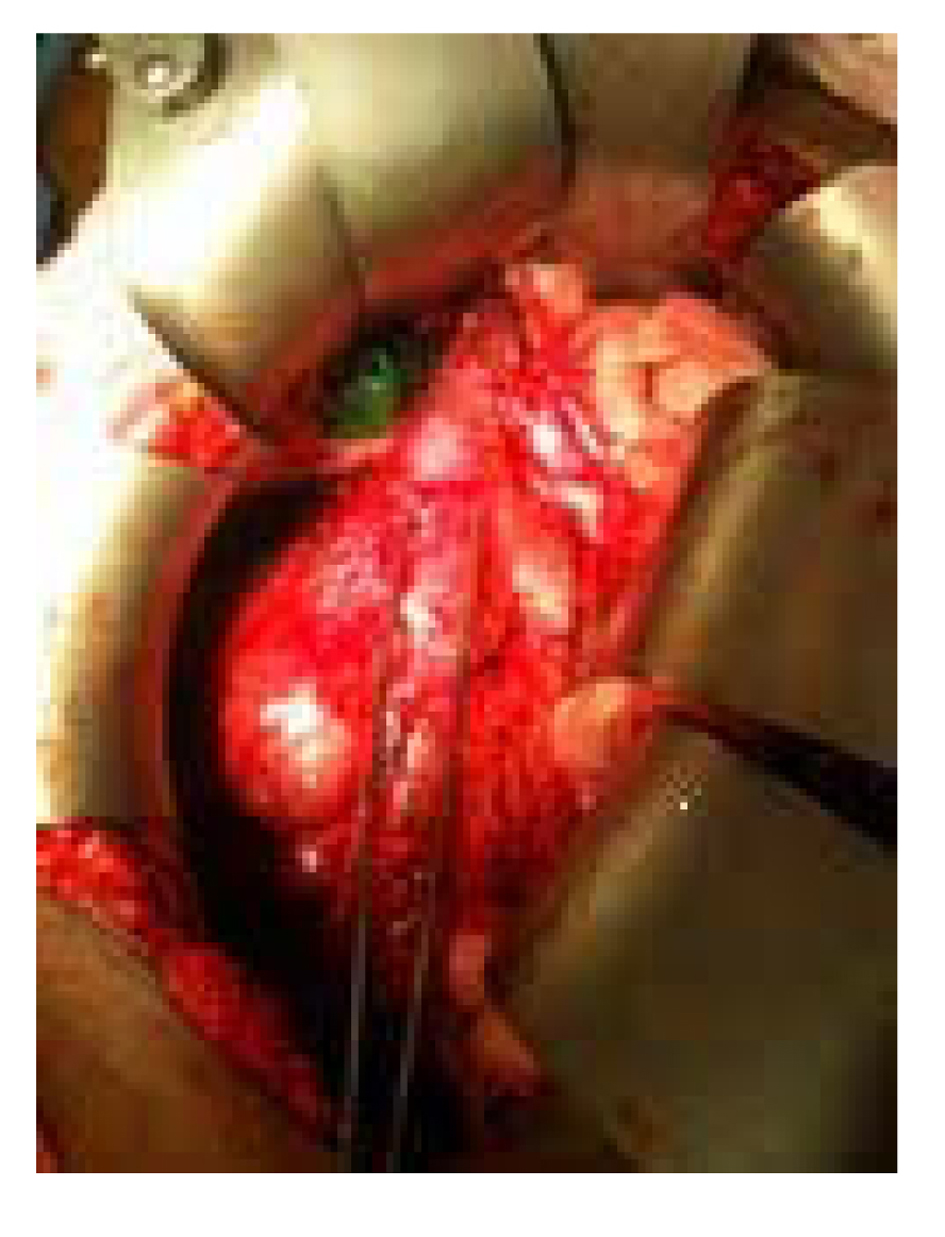
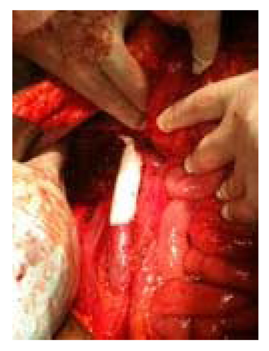
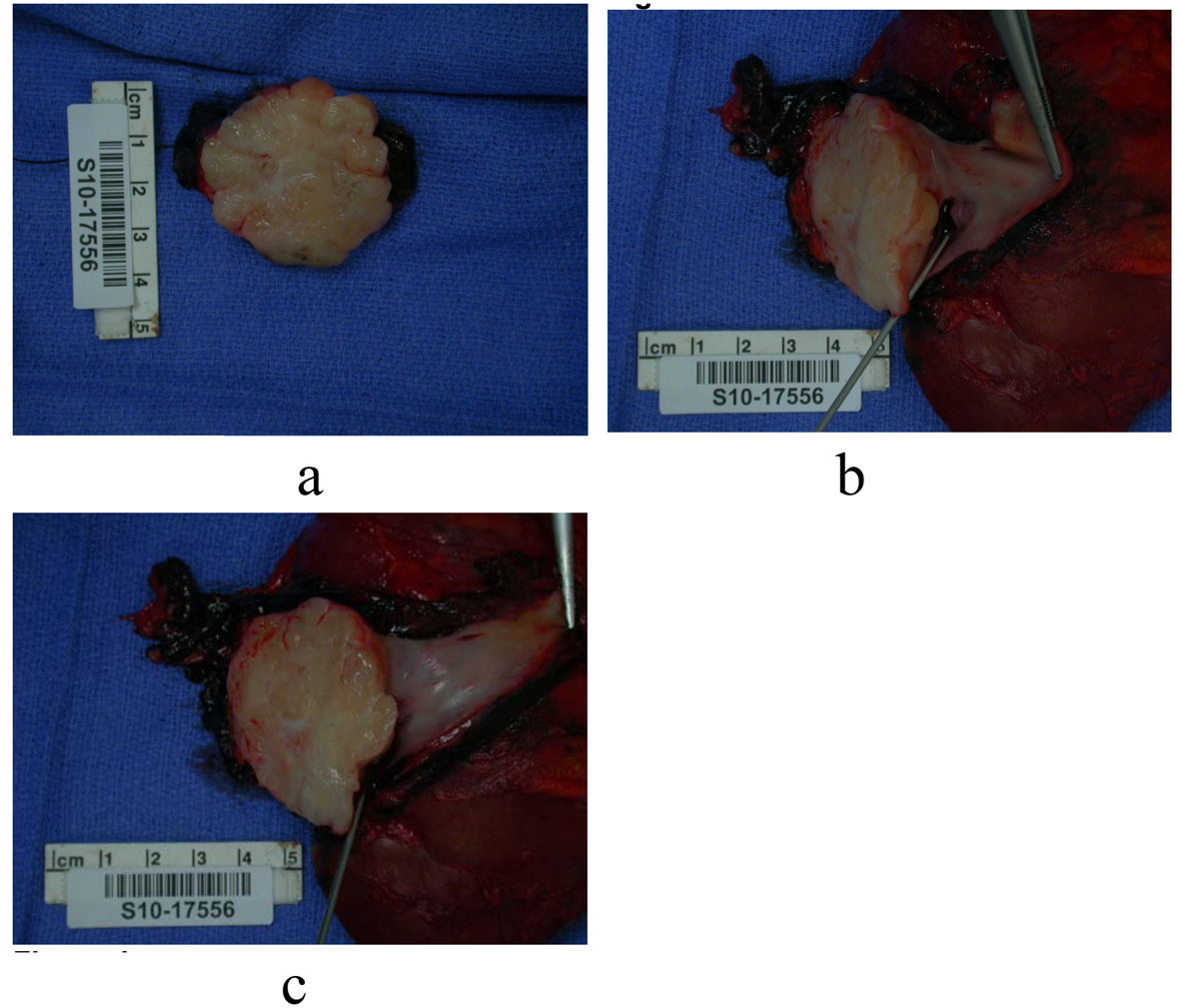
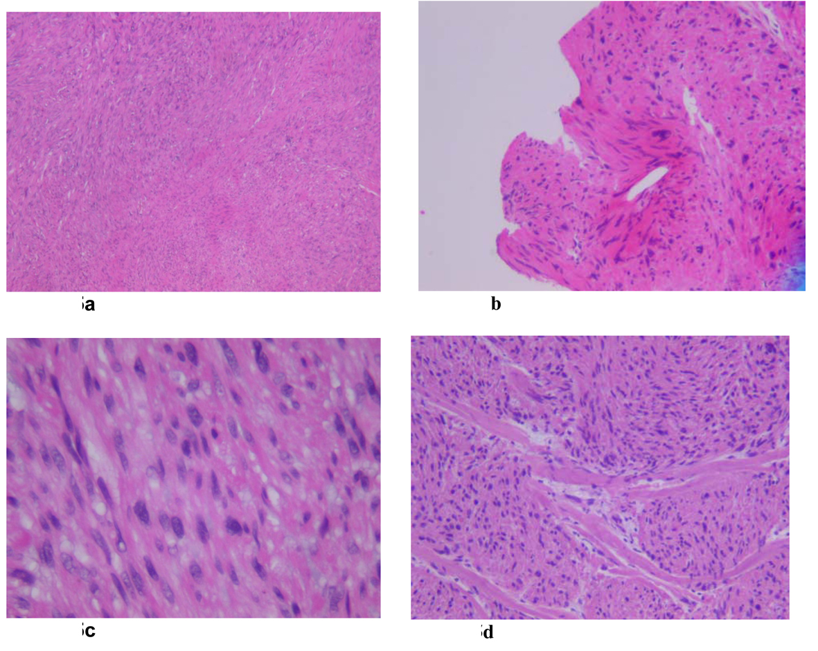
Figures