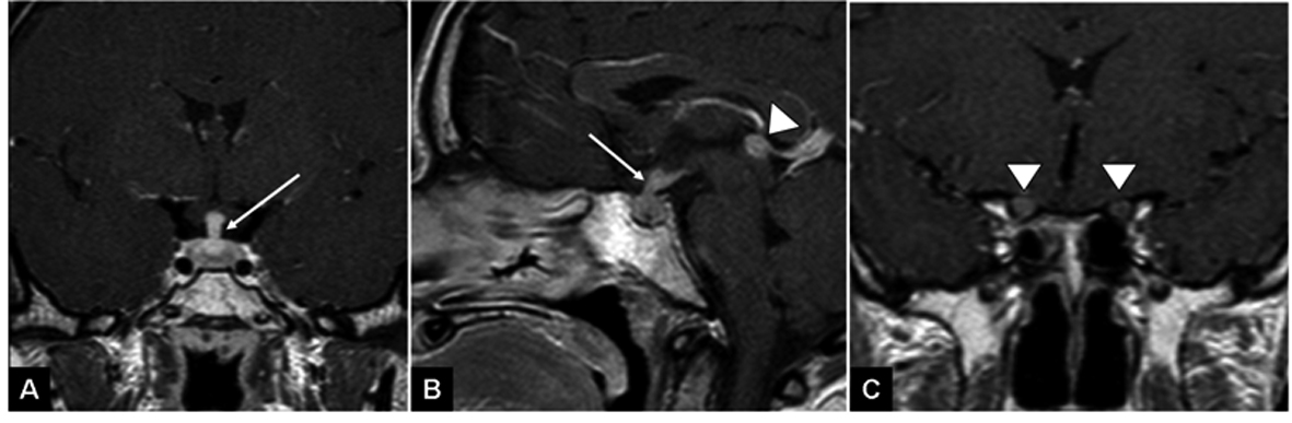
Figure 1. T1 weighted images following gadolinium injection demonstrate thickened pituitary infundibulum (arrow A, B), thickened and enhancing optic nerves (arrowheads C) and a nodular enhancing pineal lesion (arrowhead B).
| World Journal of Oncology, ISSN 1920-4531 print, 1920-454X online, Open Access |
| Article copyright, the authors; Journal compilation copyright, World J Oncol and Elmer Press Inc |
| Journal website http://www.wjon.org |
Case Report
Volume 3, Number 6, December 2012, pages 288-290
Suspected CNS Metastases of Askin's Tumor: Would You Irradiate the Neural Axis?
Figures