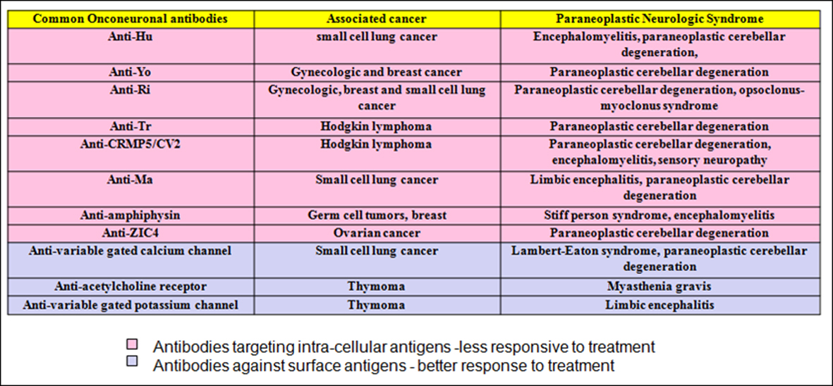
Figure 1. Common onconeuronal antibodies with their corresponding malignancies and paraneoplastic neurological syndromes.
| World Journal of Oncology, ISSN 1920-4531 print, 1920-454X online, Open Access |
| Article copyright, the authors; Journal compilation copyright, World J Oncol and Elmer Press Inc |
| Journal website http://www.wjon.org |
Case Report
Volume 3, Number 5, October 2012, pages 243-246
Diagnostic Approach to a Patient With Paraneoplastic Neurological Syndrome
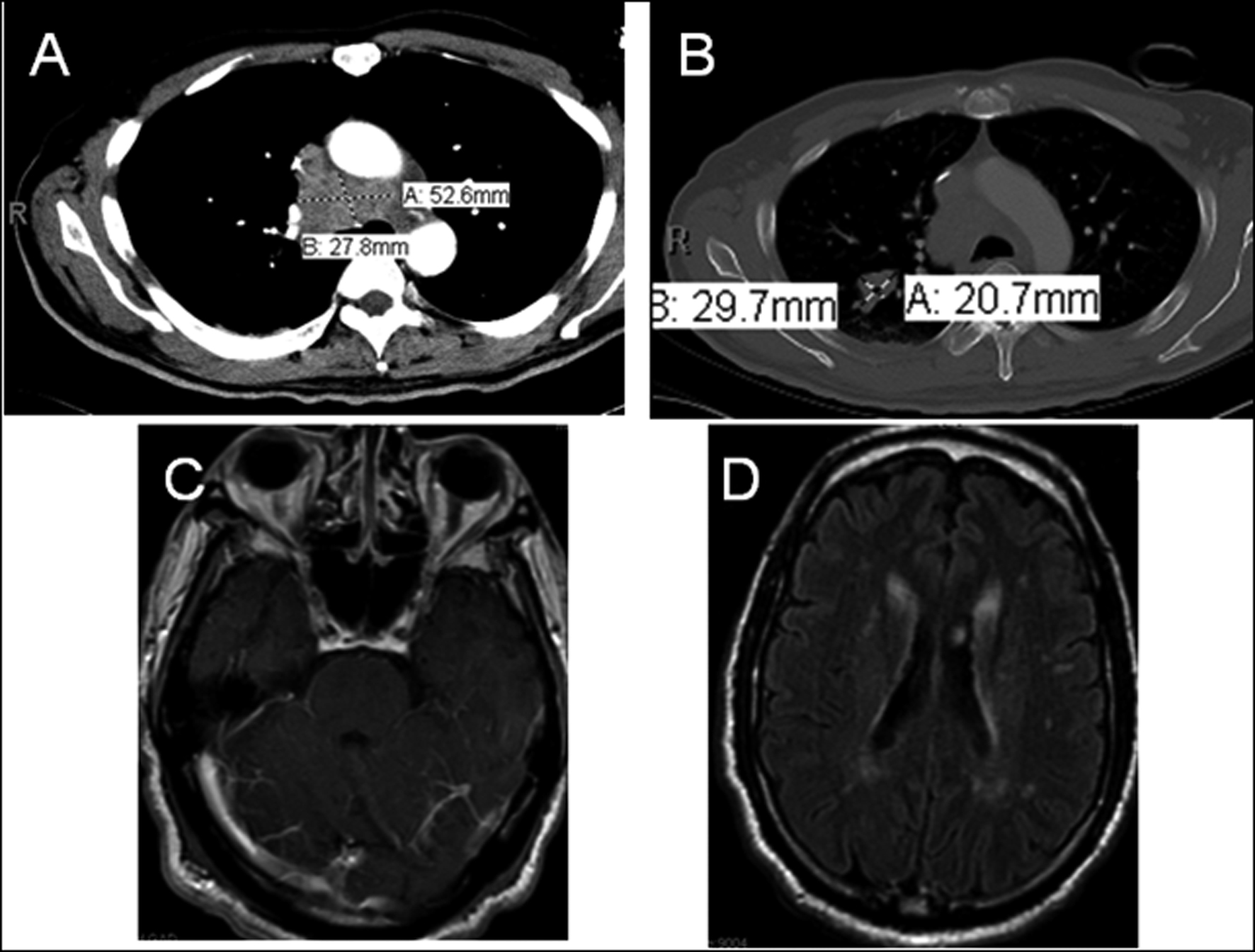
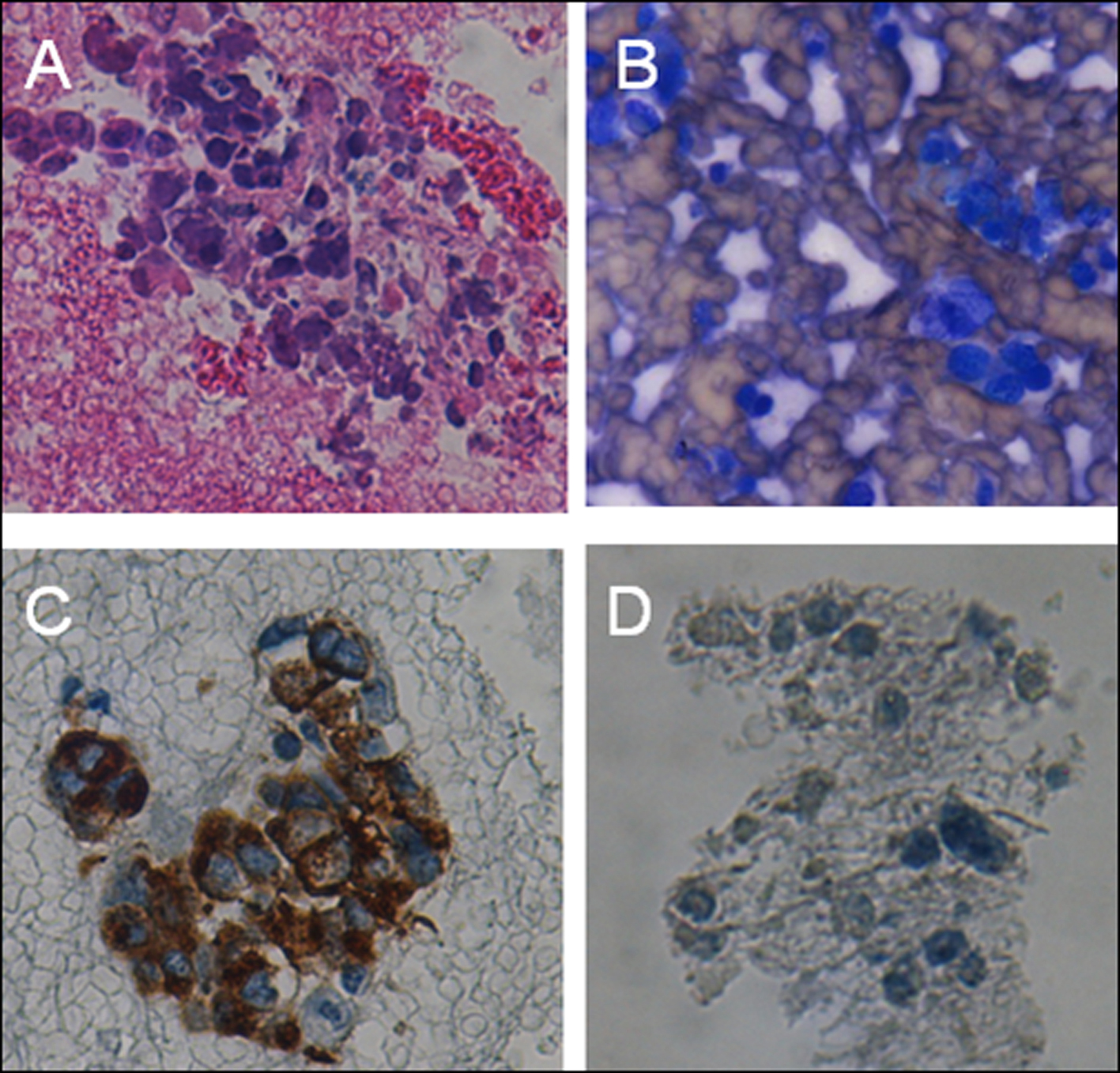
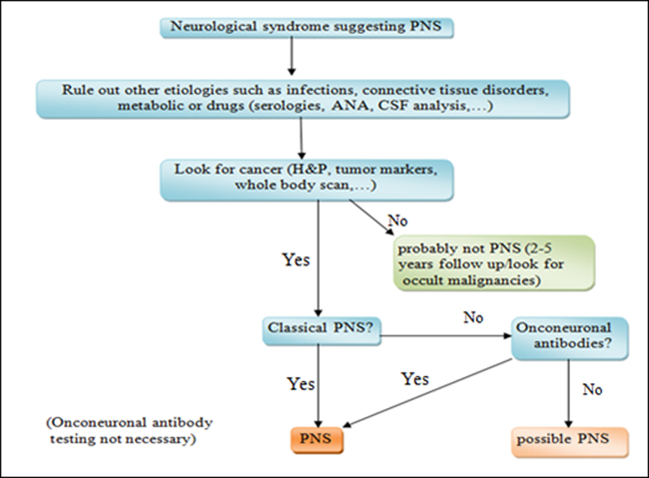
Figures