World Journal of Oncology, ISSN 1920-4531 print, 1920-454X online, Open Access
Article copyright, the authors; Journal compilation copyright, World J Oncol and Elmer Press Inc
Journal website http://www.wjon.org

Case Report

Volume 4, Number 1, February 2013, pages 54-57


Rectal Plasmablastic Lymphoma in HIV/AIDS: Two Cases

Figures

Figure 1.
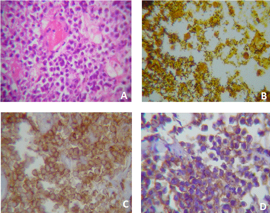
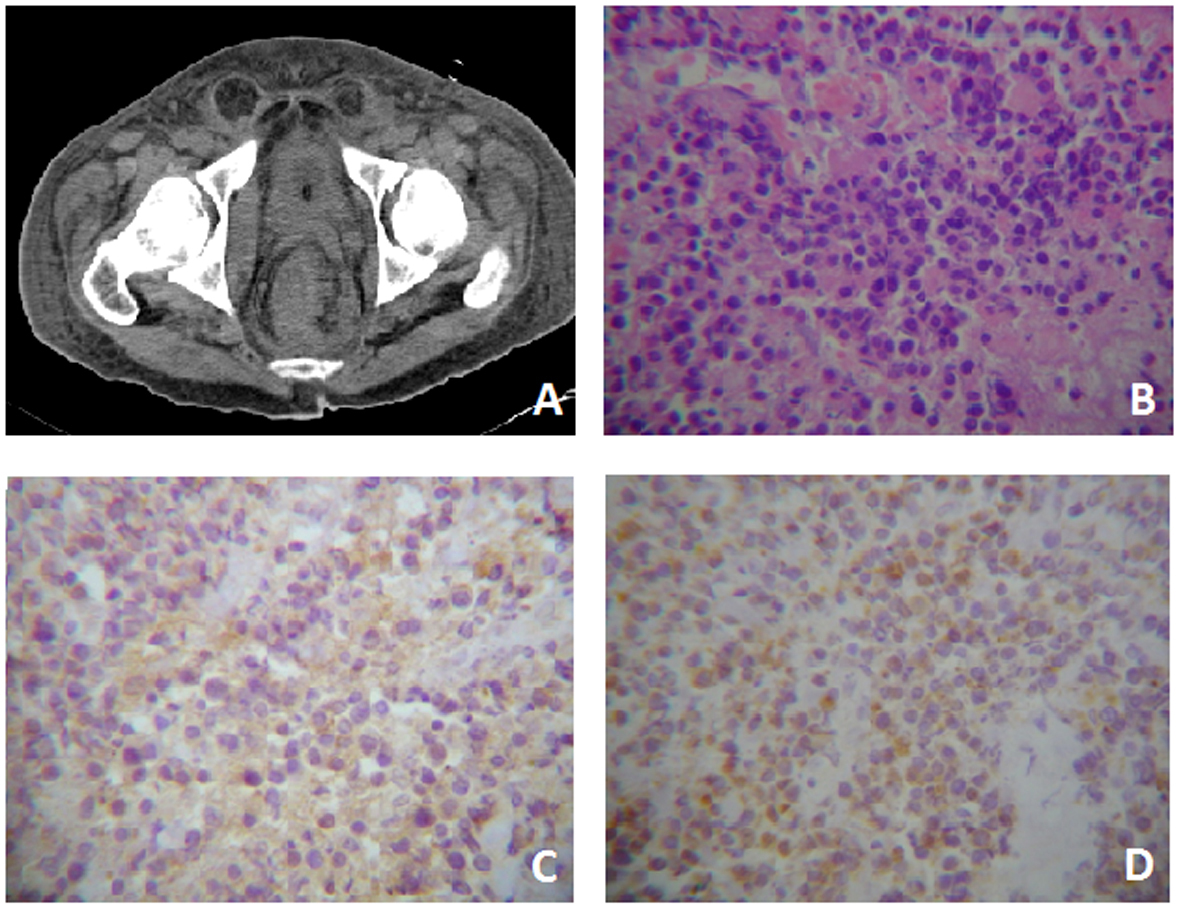
Figure 1. Case One. A) Pelvis CT where evidences rectal tumor with perirectal fascia engrossed. B) Tumor with extensive necrosis in the right lower quadrant, viable zones of monotonous, slightly cohesive, moderately pleomorphic plamacytoids showing a paranuclear light halo. HE (400 ×). C) Immunreactivity for CD-20 cytoplasmatic and membranous (400 ×). D) Immunohistochemistry for CD-138 membranous and cytoplasmatic (400 ×).
Figure 2.
Figure 2. Case two. A) Plasmacytoid cells with eccentric nucleus and a light paranuclear zone. Cells are slightly cohesive and some nucleus are wheel-shaped. It is also evident the presence of a fibrin microthrombi. HE (400X). B) Abundant bacillus colonization. Bacteria are black stained. Whartin Starry (100X). C) Membrane immunoreactivity to CD-45 (400X). D) Membrane an cytoplasmic immunoreactivity to CD-138.