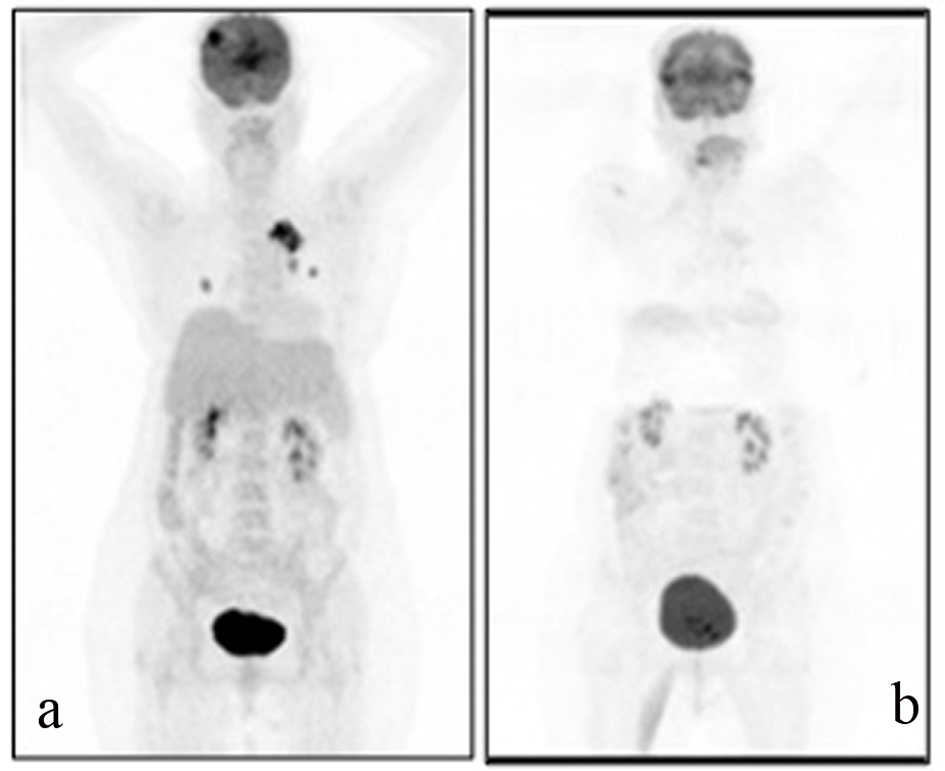
Figure 1. A 69-year-old female patient with a history of breast cancer underwent FDG PET/CT imaging before and after the therapy. For the first PET/CT imaging patient was intravenously injected 592 MBq (16 mCi) F-18 FDG after 6 hours of fasting period. Then one hour of waiting time in a silent room patient was imaged using an integrated PET/CT camera, which was consisted of a 6-slice CT gantry integrated on a LSO based full-ring PET scanner (Siements Biograph 6, IL, USA). Anterior-posterior maximum intensity projection (MIP) PET image (a) sowed intense hypermetabolic multiple nodular lesions in the right lung superior lobe with a maximum standard uptake value (SUV max) of 14.0 and left hilar hypermetabolic lymph node with a SUVmax of 6.4. In cranial slices intense hypermetabolic lesion in the right frontal lobe with a SUVmax of 13.5 was seen. Six months after the first PET/CT imaging this patient referred to Nuclear Medicine department for another PET/CT imaging after the therapy to evaluate the therapy response. Patient wan injected 418.1 MBq (11.3 mCi) F-18 FDG for imaging. Anterior and posterior MIP image (b) showed significant regression in the cranial, lung and left hilar lesions.