World Journal of Oncology, ISSN 1920-4531 print, 1920-454X online, Open Access
Article copyright, the authors; Journal compilation copyright, World J Oncol and Elmer Press Inc
Journal website http://www.wjon.org

Case Report

Volume 4, Number 2, April 2013, pages 95-101


ALK+ Anaplastic Large Cell Lymphoma With Bladder Involvement Presenting as Fever of Unknown Origin: A Case Report and Literature Review

Figures

Figure 1.
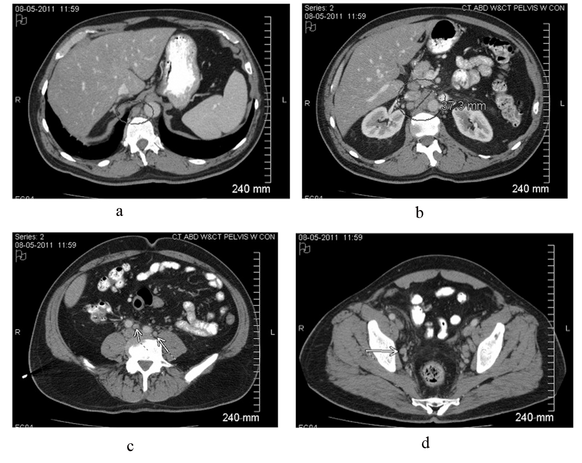
Figure 1. Contrast-enhanced CT scan of the abdomen and pelvis reveals intrabdominal and retroperitoneal adenopathy of uncertain origin. Adenopathy includes right retrocrural, retroperitoneal, celiac, peripancreatic, mesenteric, iliac and pelvic lymph nodes. Upper retroperitoneum reveals confluent nodal mass measuring 37.3 millimeters in image b.
Figure 2.
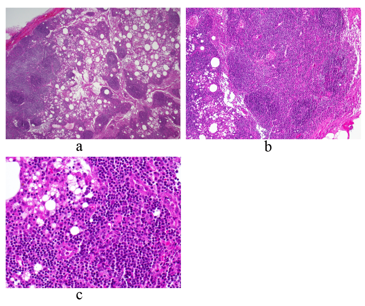
Figure 2. Periaortic lymph node biopsy reveals a mature and polymorphous lymphoid population with some macrophages and normal histologic architecture. There is an intact capsule and no lymphocyte migration outside of the capsule. There are follicles with germinal centers and normal interfollicular cellularity. Photos a, b and c represent low, medium and high power views, respectively.
Figure 3.
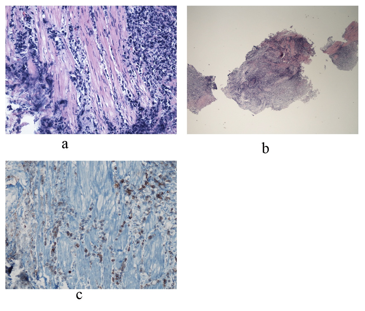
Figure 3. Bladder biopsy sections reveal a dense infiltrate composed of large abnormal lymphoid cells at low and medium power views. Photo a shows diffuse, ragged infiltration of bladder muscle by cells with dark nuclei and moderate size. An example of these cells is represented in photo b. Immunohistochemical stains showed that these large cells are positive for CD45, CD3, CD5, CD4, and CD30. CD30 positive cells are represented in photo c. Occasional large cells are also positive for ALK-1. They are negative for CD20, CD8, CD34, Tdt, CD56, pankeratin, synaptophysin and chromogranin. Findings are consistent with an ALK+ anaplastic large cell lymphoma.

Tables

Table 1. Diagnostic Laboratory Work-Up Performed at Initial Hospital Admission
 
LabResultLabResult
Wnl: within normal limits; Neg: negative. ESR: erythrocyte sedimentation rate; CRP: C-reactive protein; C3/C4: complement; RF: rheumatoid factor; LDH: lactate dehydrogenase; ANA: anti-nuclear antigen; dsDNA: double stranded DNA; SS-a/b: Sjogren’s syndrome; Scl: scleroderma; RNP: ribonucleoprotein; Sm: Smith; C/P-ANCA: cytoplasmic/perinuclear antineutrophil cytoplasmic antibodies; TSH: thyroid stimulating hormone; T4: thyroid hormone; SPEP: serum protein electrophoresis; AFP: alpha-fetoprotein; B-HCG: beta-human chorionic gonadotropin; PPD: purified protein derivative; ASO: anti-streptolysin titer; Ig: immunoglobulin; PCR: polymerase chain reaction; EBV: Ebstein-barr virus; HIV: human immunodeficiency virus.
InflammatoryESR65 (H)InfectiousPPDNeg
CRP12.9 (H)ASONeg
C3132Hepatitis panelNeg
C430Legionella urinary antigenNeg
RF< 20Parvovirus IgGWnl
LDH315 (H)Parvovirus IgM
AntibodiesANA+ 1:160Coccidiodes antibodyNeg
dsDNAWnlCMV IgGWnl
SS-a/SS-bNegCMV IgMWnl
Scl-70NegCMV PCRUndetectable
RNPNegEBV IgG5.05 (H)
SmNegEBV IgM0.01
Sm/RNPNegEBV PCRUndetectable
C-ANCA/P-ANCA< 1:20Lyme Disease PCRUndetectable
EndocrineTSH1.42Malarial smearNeg
Free T41.8HIV screenNeg
A.M.-Cortisol27Histoplasmosis urinary antigenNeg
SPEPNo monoclonal proteinBlastomycosis urinary antigenNeg
AFP1.7Blood cultures × 6No growth
B-HCG1.7Urine cultures × 2No growth


Table 2. Diagnostic Imaging Performed at Initial Hospital Admission
 
ImageResult
Neg: negative; LVSF: left ventricular systolic function; EF: ejection fraction; LVH: left ventricular hypertrophy; MR: mitral regurgitation; TR: tricuspid regurgitation; RVSP: right ventricular systolic pressure; HTN: hypertension.
CT chest w/IV contrastNeg. for mass or lymphadenopathy. Lungs clear.
Renal UltrasoundBilateral complex renal cysts: 2.9 cm in right kidney and 1.8 cm in the left kidney.
Transthoracic Echocardiogram- Normal cardiac chamber size. Normal LVSF, EF 60%. Mild concentric LVH.
- Trivial MR. Mild TR, estimated RVSP of 42 mmHg suggesting mild pulmonary HTN.
- Small circumferential pericardial effusion w/o evidence of tamponade.
Nuclear Medicine Bone ScanNonspecific uptake in the right superolateral bony orbit.
Bilateral Lower Extremity Venous DopplerNegative for deep venous thrombosis.