World Journal of Oncology, ISSN 1920-4531 print, 1920-454X online, Open Access
Article copyright, the authors; Journal compilation copyright, World J Oncol and Elmer Press Inc
Journal website http://www.wjon.org

Case Report

Volume 4, Number 3, June 2013, pages 169-172


Renal Medullary Carcinoma Masquerading as Bilateral Breast Carcinoma Category: Case Report

Figures

Figure 1.
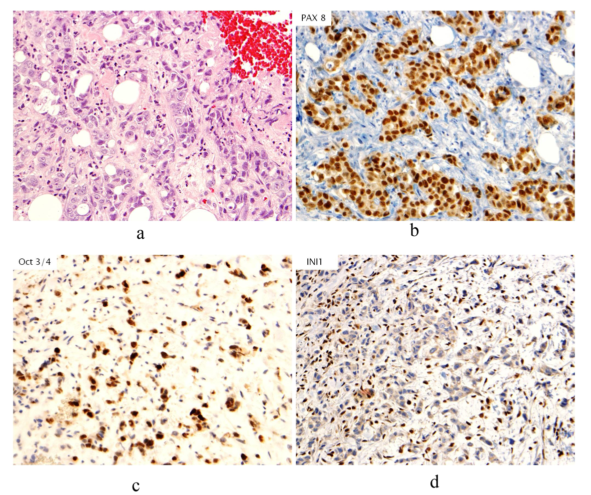
Figure 1. (a). Hematoxylin & Eosin stain (H&E stain) of biopsied breast tissue showing infiltrative, poorly differentiated carcinoma growing in cords and forming occasional tubules. No associated ductal carcinoma in situ component, microcalcifications or lymphovascular involvement by tumor is seen; (b). Immunohistochemical stain of PAX8 revealing nuclear staining in neoplastic cells; (c). Immunohistochemical stain of Oct3/4 revealing nuclear staining in neoplastic cells; (d). Immunohistochemical stain of INI1 showing absence of staining in neoplastic cells.
Figure 2.
Figure 2. MRI breast (T1, fat saturated, post-contrast subtraction image) showing the largest right breast mass (left arrow) which is heterogeneously enhancing, with central necrosis and the largest left breast mass (right arrow), also heterogeneously enhancing with central necrosis.
Figure 3.
Figure 3. Axial CT (with oral and intravenous contrast enhancement) showing a 6.7 × 6.9 cm heterogeneous exophytic mass with areas of central low attenuation arising from the lower pole of the right kidney, with areas of central necrosis.
Figure 4.
Figure 4. Coronal CT (with oral and intravenous contrast enhancement) showing large heterogeneously enhancing mass replacing lower pole of the right kidney.