World Journal of Oncology, ISSN 1920-4531 print, 1920-454X online, Open Access
Article copyright, the authors; Journal compilation copyright, World J Oncol and Elmer Press Inc
Journal website http://www.wjon.org

Case Report

Volume 5, Number 4, August 2014, pages 183-186


Another Case of Pulmonary Edema or May Be Not: An Unusual Presentation of Metastatic Melanoma

Figures

Figure 1.
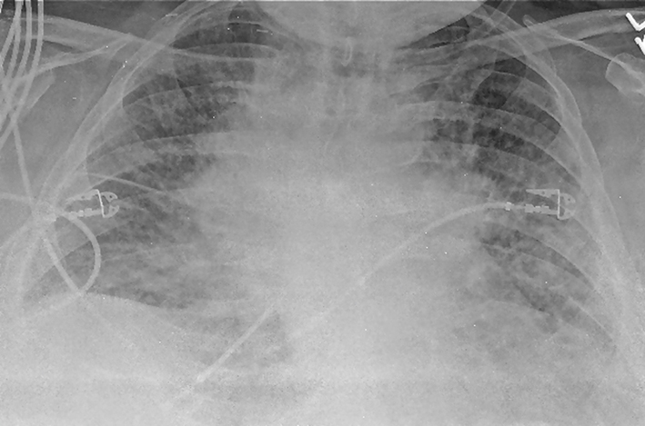
Figure 1. Chest X-ray on admission showed bilateral perihilar haziness and fluid in right transverse fissure consistent with pulmonary edema.
Figure 2.
Figure 2. A repeat chest X-ray (day 7) revealed diffuse bilateral infiltrates or edema, predominantly interstitial in character with cardiomegaly (not significantly improved from admission, in spite of aggressive diuresis).
Figure 3.
Figure 3. Patient’s CT scan of chest on day 7 (A, B) demonstrated mediastinal lymphadenopathy, diffuses pulmonary nodules, multifocal consolidations in right upper and lower lobe, lingular lobe, and left lower lobe as well as bilateral pleural effusion.
Figure 4.
Figure 4. Histopathology of transbronchial biopsy from right upper lobe: (A) × 100 magnification and (B) × 400 magnification, showing malignant epithelioid cells with immunohistochemical staining positive for S100, Melan A and MITF, morphologically consistent with metastatic melanoma.