World Journal of Oncology, ISSN 1920-4531 print, 1920-454X online, Open Access
Article copyright, the authors; Journal compilation copyright, World J Oncol and Elmer Press Inc
Journal website http://www.wjon.org

Case Report

Volume 6, Number 1, February 2015, pages 297-300


Obstructive Jaundice as an Uncommon Manifestation of Metastatic Breast Cancer

Figures

Figure 1.
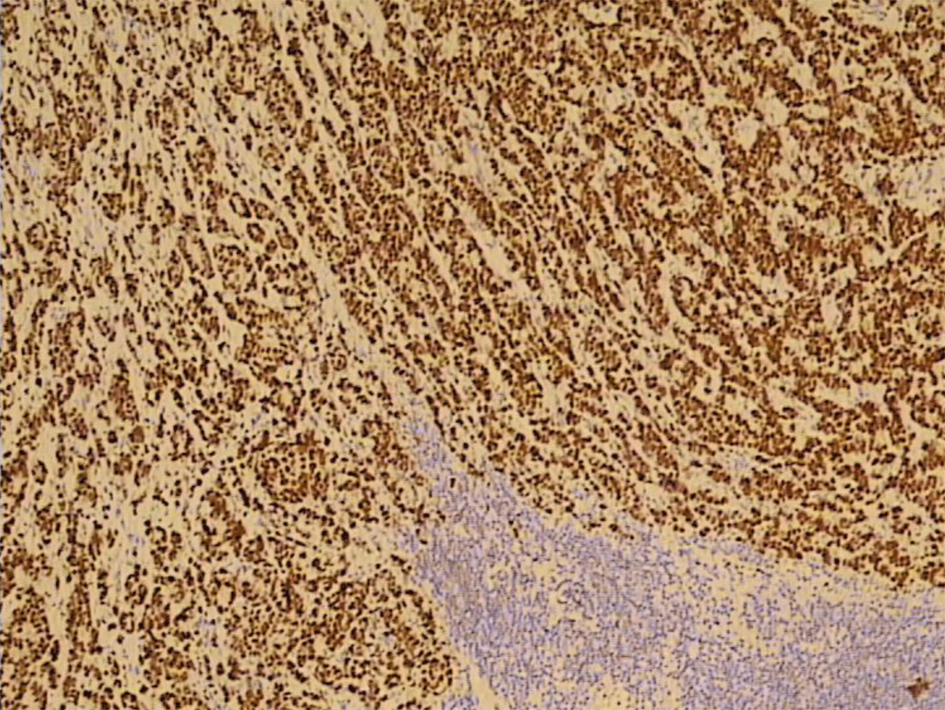
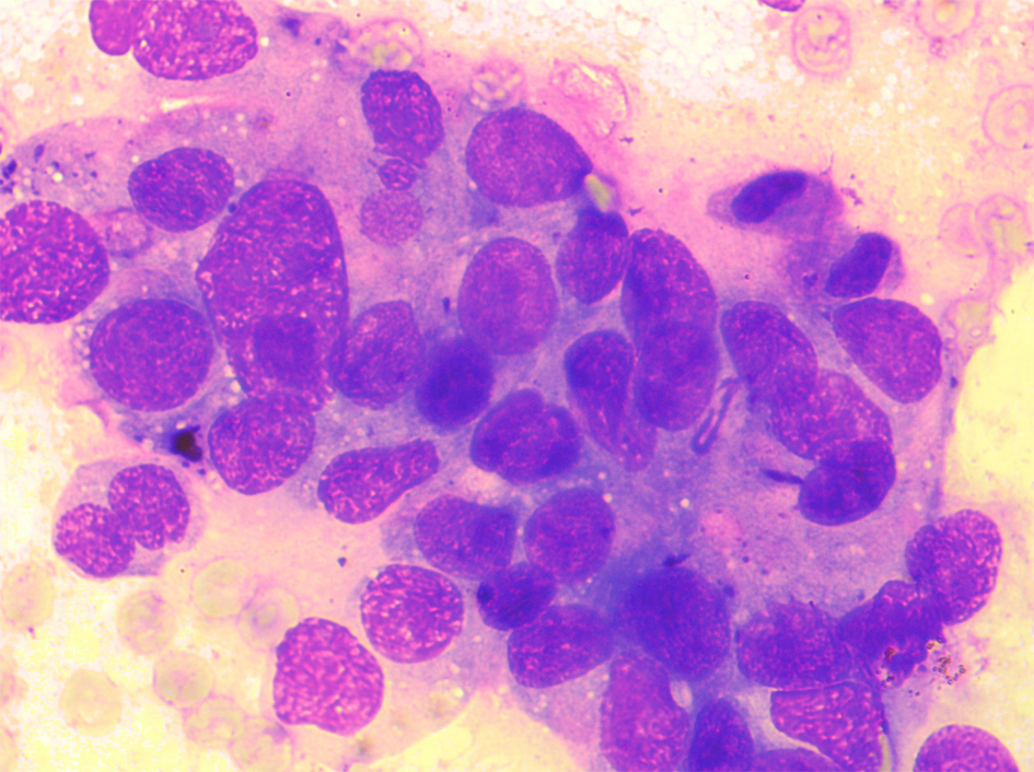
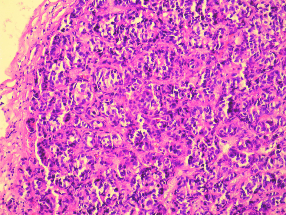
Figure 1. Histological analysis of the extirpated lymph nodes. Hemalaun-eosin stain showing infiltration with tubular formations and clusters of atypical epithelial cells which infiltrated the lymph node’s capsule (HE, × 20).
Figure 2.
Figure 2. A strong positive nuclear reaction to estrogen receptor in 100% of tumor cells (IMH, × 10).
Figure 3.
Figure 3. Immunohistochemically, moderate-to-strong nuclear reaction to progesterone receptor in 60% of tumor cells (IMH, × 20).
Figure 4.
Figure 4. ERCP showing a subtotal stenosis of the common bile duct 3 cm in length with expressed prestenotic dilatation of both hepatic ducts and intrahepatic bile ducts.
Figure 5.
Figure 5. ERCP showing an implanted plastic stent with adequate position in the common bile duct and appropriate function.
Figure 6.
Figure 6. Cytological analysis of the material obtained during ERCP. The smears are showing several smaller and a few larger, often three-dimensional groups of partially degenerative, well-to-moderately differentiated malignant cells, originating from glandular epithelium (MGG, × 40).