World Journal of Oncology, ISSN 1920-4531 print, 1920-454X online, Open Access
Article copyright, the authors; Journal compilation copyright, World J Oncol and Elmer Press Inc
Journal website http://www.wjon.org

Case Report

Volume 5, Number 3, June 2014, pages 139-143


Extraconal Orbital Soft Tissue Metastasis Secondary to Prostate Cancer: An Unusual Presentation

Figures

Figure 1.
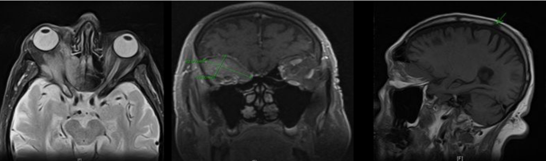
Figure 1. MRI of the head showing a right intra-orbital mass measuring 4.3 × 2.3 cm, extending into the anterior cranial fossa (superiorly); lamina papyracea, posterior ethmoid sinus and anterior lateral sphenoid sinus (medially); and posterior lateral orbital wall and sphenoid wing (laterally); sclerotic lesion in the right parietal bone also noted.
Figure 2.
Figure 2. Whole body PET-CT showing FDG avid uptake in posterior lobe of prostate, axial spine, iliac bones and bilateral iliac lymph nodes.
Figure 3.
Figure 3. MRI of the spine showing multi-level osseous metastatic infiltration throughout the spinal axis particularly involving the lower thoracic and lumbar regions.
Figure 4.
Figure 4. Lesions arising in the extraconal space.

Table

Table 1. Case Reports of Uveal Tract Metastasis Secondary to Prostate Cancer
 
AuthorAge (years)YearSite of orbital metastasis
Sarenac et al [13]692012Bilateral iris metastasis
Keizur et al [14]651995Choroidal metastasis
Liu et al [15]601992Combined scleroidal and choroidal metastasis
Dieckert et al [16]541982Choroidal metastasis
Zappia et al [17]581972Metastasis to both the choroid and optic nerve, mimicking papilledema