World Journal of Oncology, ISSN 1920-4531 print, 1920-454X online, Open Access
Article copyright, the authors; Journal compilation copyright, World J Oncol and Elmer Press Inc
Journal website http://www.wjon.org

Case Report

Volume 5, Number 3, June 2014, pages 149-152


Pulmonary Embolism as an Initial Presentation of Adrenocortical Carcinoma

Figures

Figure 1.
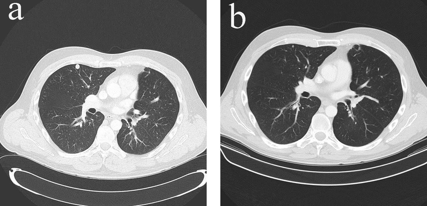
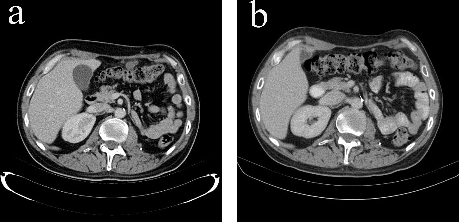
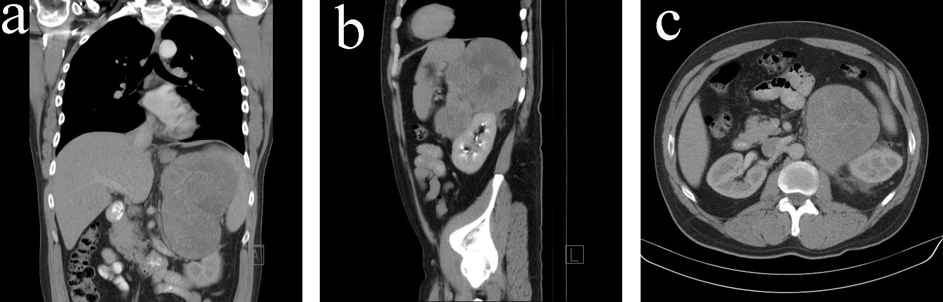
Figure 1. (a) CT scan (coronal view) on initial diagnosis. (b) CT scan (sagittal view) on initial diagnosis. (c) CT scan (transverse view) on initial diagnosis.
Figure 2.
Figure 2. Macroscopic appearance of the resected tumor.
Figure 3.
Figure 3. (a) Hematoxylin and eosin stain. (b) Vimentin stain. (c) Melan-A stain.
Figure 4.
Figure 4. (a) Pulmonary metastases in CT scan (transverse view) before additional chemotherapy with EDP. (b) Resolution of pulmonary metastases in CT scan (transverse view) after additional chemotherapy with EDP.
Figure 5.
Figure 5. (a) Peritoneal metastases in CT scan (transverse view) before additional chemotherapy with EDP. (b) Resolution of peritoneal metastases in CT scan (transverse view) after additional chemotherapy with EDP.