World Journal of Oncology, ISSN 1920-4531 print, 1920-454X online, Open Access
Article copyright, the authors; Journal compilation copyright, World J Oncol and Elmer Press Inc
Journal website http://www.wjon.org

Case Report

Volume 5, Number 5-6, December 2014, pages 237-239


Pulsed Erlotinib as Sole Treatment of Leptomeningeal Carcinomatosis: Can We Avoid the Use of Intrathecal Therapy?

Figures

Figure 1.
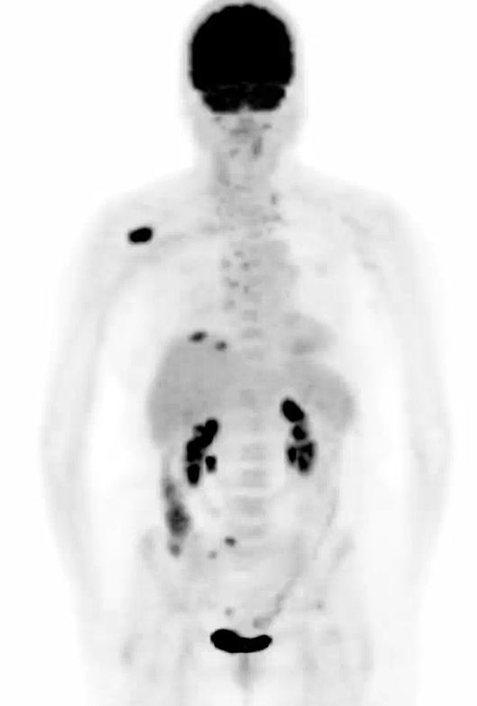
Figure 1. Computed tomography-positron emission tomography performed prior to treatment. Maximum intensity projection image shows mass within right lung base. Adenopathy with abnormal uptake is present throughout the mediastinum and neck. Largest bone metastasis involves right scapula.
Figure 2.
Figure 2. Leptomeningeal carcinomatosis. (A) Brain magnetic resonance imaging (MRI) obtained when patient first presented with neurologic deficits shows multiple sites of leptomeningeal enhancement on post-contrast T1-weighted sequences (arrows). (B) Following pulsed-dose erlotinib therapy, repeat MRI scan showed complete resolution of tumor.