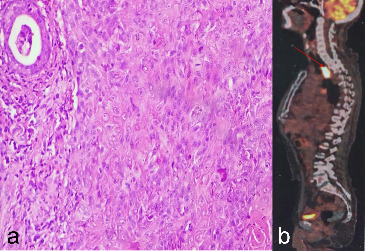
Figure 1. (a) Moderately differentiated squamous cell carcinoma. (b) PET-CECT showing FDG avid mass lesion in esophagus at level of D2-D4.
| World Journal of Oncology, ISSN 1920-4531 print, 1920-454X online, Open Access |
| Article copyright, the authors; Journal compilation copyright, World J Oncol and Elmer Press Inc |
| Journal website http://www.wjon.org |
Case Report
Volume 7, Number 5-6, December 2016, pages 119-123
Radiation-Induced Malignancies: Our Experiences With Five Cases
Figures
Table
| Case | Primary diagnosis | Histology of primary | Radiotherapy dose | Chemotherapy agent | Latent period (years) | Site of RIM | Histology of RIM | Treatment of RIM |
|---|---|---|---|---|---|---|---|---|
| IDC: infiltrating ductal carcinoma; CAF: cyclophosphamide, adriamycin, 5-florouracil; E: esophagus; MDSCC: moderately differentiated squamous cell carcinoma; RT: radiotherapy; U: uterus; CS: carcinosarcoma; CT: chemotherapy; BOT: base of tongue; NEC: neuroendocrine carcinoma; BM: buccal mucosa; L: leiomyosarcoma; PDSCC: poorly differentiated squamous cell carcinoma; GBS: gingivobuccal sulcus; CCRT: concurrent chemoradiotherapy. | ||||||||
| 1 | Carcinoma breast | IDC | 50 Gy in 25 fractions | Six cycles of CAF regimen | 14 | E | MDSCC | Palliative RT |
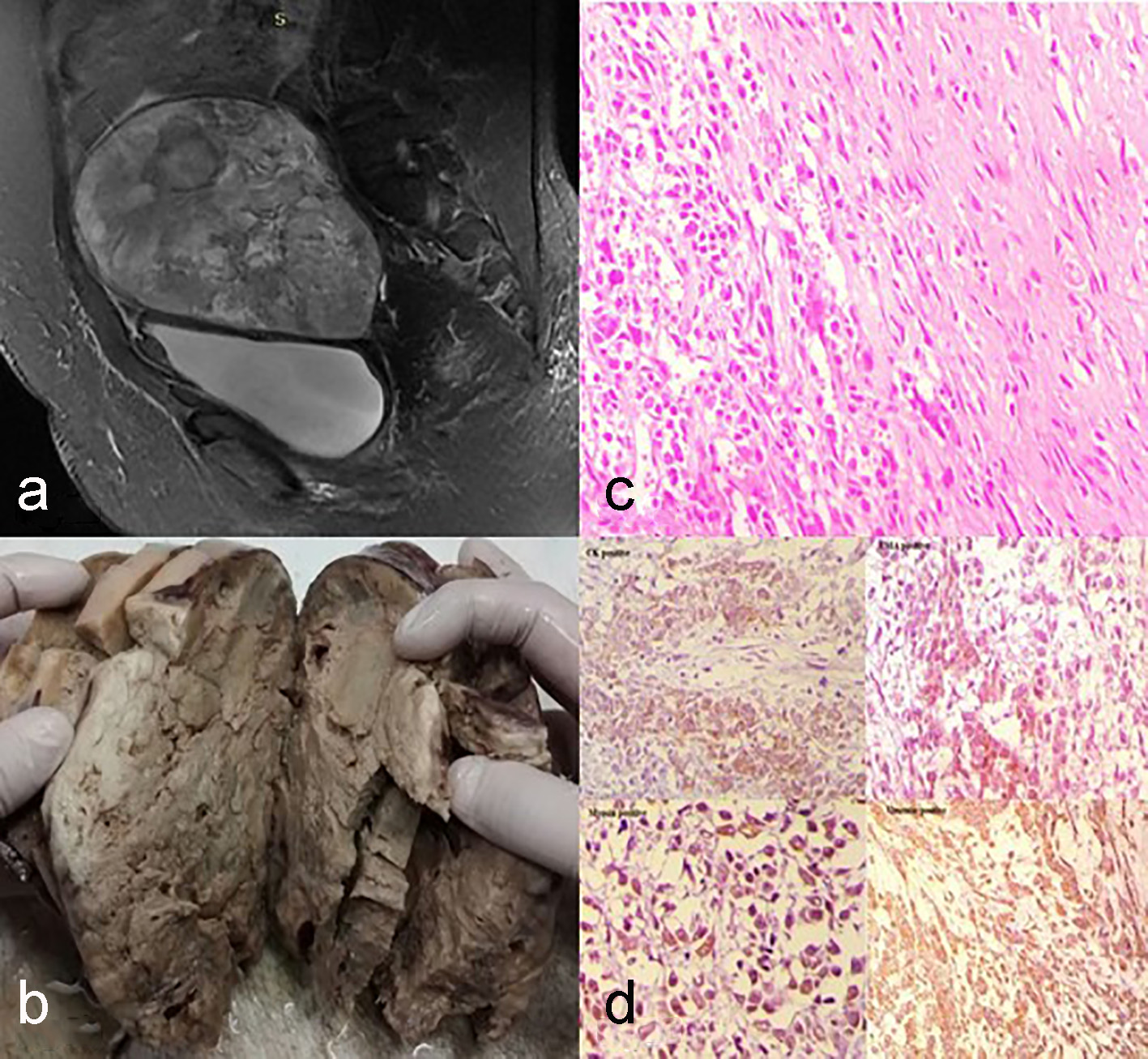
| 2 | Carcinoma cervix | MDSCC | 50 Gy in 25 fractions f/b 7 Gy in 3 fractions | Cisplatin | 13 | U | CS | CT |
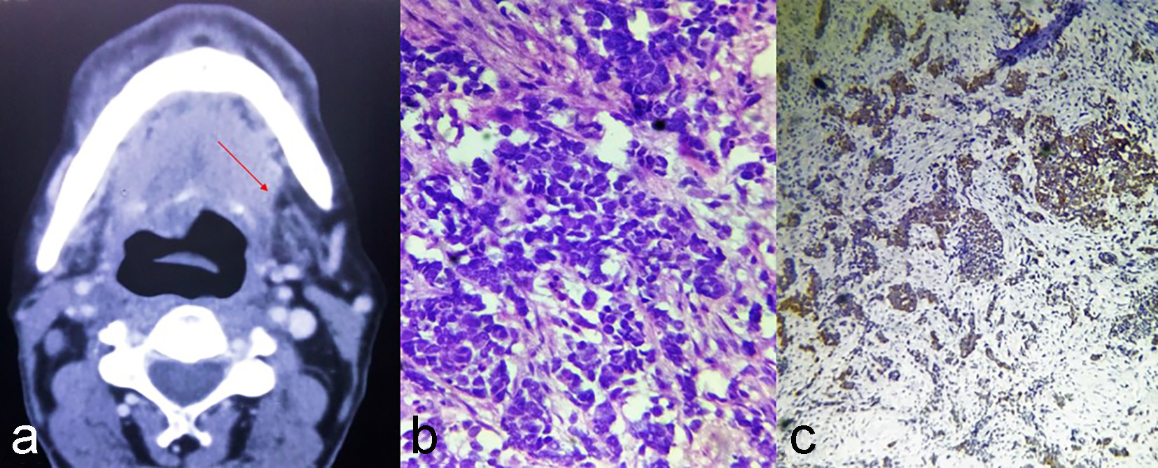
| 3 | Carcinoma BOT | MDSCC | 66 Gy in 33 fractions | Cisplatin | 7 | BOT | NEC | CT |
| 4 | Carcinoma BM | WDSCC | 66 Gy in 33 fractions | Cisplatin | 4 | BM | L | Surgery |
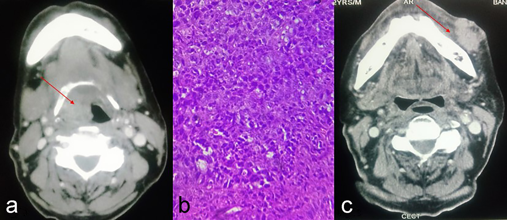
| 5 | Carcinoma supraglottis | PDSCC | 66 Gy in 33 fractions | Cisplatin | 3 | GBS | MDSCC | CCRT |